Current Trends To Deal Endodontic Emergencies and Use of Antibiotics by Dentists of Karachi

Sara Ikram Khan1                             BDS

Shama Asghar2                                  BDS, FCPS

Syed Muhammad Faizan3                BDS

INTRODUCTION:

The aim of this study was to collect information from dentists regarding treatment of endodontic emergencies and investigate their antibiotic prescribing habits.

METHODOLOGY: A cross sectional study was conducted in which questionnaires were distributed to 300 dentists of dental hospitals and private clinics of Karachi, Pakistan. The survey dealt with questions focusing on treatment approaches for different endodontic emergency situations and antibiotic prescribing habits of the dentists.

RESULTS: A total of 182 participants were included with a response rate of 65%. In cases of a vital teeth with irreversible pulpitis, (59.9%) of dentists opted for two visit root canal treatment. Dentists with working experience 5 years performed root canal treatment in more than two visits which is in a higher ratio than the other groups (P<0.05). (75.3%) of dentists prescribed medications if RCT required multiple visits. (33.5%) managed emergency case of pulpitis by pulpectomy in combination with analgesics and antibiotics. In management of acute apical periodontitis, (28%) opted for pulpectomy with intra-canal medication. In case of acute apical abscess, (67.6%) of the dentists preferred drainage of the abscess by opening pulp chamber in combination with antibiotics. Most frequently prescribed antibiotics were Amoxicillin-Clavulanate (78.8%).

CONCLUSIONS: Dentists were over prescribing antibiotics in may conditions. Educational programs should be conducted on regular basis to increase the knowledge of the dentists.

KEY WORDS: Endodontic emergencies, Root Canal Treatment, Antibiotic prescription, Dentists.

HOW TO CITE:  Khan SI, Asghar S, Faizan SM. Current trends to deal endodontic emergencies and use of antibiotics by dentists of karachi. J Pak Dent Assoc 2018;27(1):18-21.

DOI: https://doi.org/10.25301/JPDA.271.18

Received: 23 November, 2017,  Accepted: 08 December, 2017

INTRODUCTION

Dental emergencies usually include severe pain, swelling and abscess formation which are dealt on daily basis by either endodontic treatment or extraction of the tooth.1 Dental emergencies are usually a result of untreated irreversible pulpitis characterized by spontaneous episodes of sharp shooting and lingering pain which is due to irreparable pulpal damage2 and if not treated can lead to pulp necrosis and abscess formation. Fractured and cracked tooth that involves pulp can also lead to similar episodes of pain3 and require RCT or extraction. These situations are further dealt with systemic antibiotics and analgesics for relief of symptoms as an adjunctive treatment if indicated.1
However, literature does not present much evidence to support use of antibiotic for pain relief4,5 and unnecessary prescription of systemic antibiotics leads to systemic side effects, allergic reactions and antibiotic resistance.6,7 Dentists need to be educated about the antibiotic prescription guidelines and should only prescribe antibiotics if it is clearly indicated.1,2,4,6,7 The aim of this study is to collect information about the knowledge and practice of the dental practitioners of Karachi regarding endodontic emergencies and also investigate their antibiotic prescribing habits.

METHODOLOGY

It was a cross-sectional, multi-center study. A two-page content validated questionnaire was prepared in English language as our study tool. The ethical approval was obtained by the ethical review committee of Bahria University Medical and Dental College. The questionnaire was pilot testing by 7 dentists each from 3 different dental hospitals and 4 private practitioners before data collection. The questionnaire was then distributed to 300 dentists of private dental clinics as well as dental hospitals associated with institutes in Karachi, Pakistan. Consent was obtained from the respondents and no names were asked to ensure anonymity.

The questionnaire was divided into two parts; the first comprised of questions regarding years of professional activity, number of appointments dentists took for treating vital and non-vital teeth, number of appointments dentists took for single rooted and multi-rooted teeth and whether they prescribed analgesics and antibiotics in between the appointments. The second part focused on different treatment approaches in various endodontic emergencies like irreversible pulpitis, acute apical periodontitis, acute apical abscess and lastly the most commonly prescribed antibiotics were asked. Data analysis was done on IBM SPSS Statistics version 20. Frequency tables were prepared and statistical analysis using chi square test was done and P value of <0.05 was considered significant.

RESULTS

Of the 300 questionnaires that were distributed, 195 dentists responded and returned the questionnaires among which 13 were excluded due to incomplete information. Over all response rate was 65% (n=182). In cases of a vital teeth with irreversible pulpitis, (59.9%) of dentists opted for two visits for root canal treatment, while (33.5%) opted for more than two visits. Dentists who had a working experience of less than 5 years performed root canal treatment in more than two visits which is in a higher ratio than the other groups (P<0.05) (Table 1). (52.7%) practitioners preferred multiple visits for treatment of a non

Table 1: Number of appointments in Root Canal Treatment of Vital and Non-Vital teeth

vital tooth. (51%) practitioners opted for a single-visit root canal treatment for incisors and canines, (63.2%) opted for double visits in case of premolars and for (76.4%) preferred three or more visits for molars. (75.3%) of dentists prescribed medications if RCT required multiple visits. (47.8%) of respondents managed emergency case of irreversible pulpitis by pulpectomy in combination with analgesics whereas (33.5%) dentists prescribed antibiotics in addition to pulpectomy and analgesics. Only (2.7%) of respondents opted for analgesics and antibiotics for this case. Rate of antibiotic prescription by dentists with working experience of 6-15 years was significantly lower than the young practitioners (P<0.05) (Table 2).

Table 2: Emergency treatment approaches for irreversible pulpitis, AAP, AAA

Cases of acute apical periodontitis (51.1%) of the respondents opted for pulpectomy in combination with analgesics and antibiotics while (28%) used pulpectomy with the use of intra-canal medication. (17.6%) of the dentists managed the case with analgesics and antibiotics.

In case of acute apical abscess, (67.6%) of the dentists opted for drainage of the abscess by opening pulp chamber in combination with antibiotics. (24.2%) dealt the case just by opening the pulp chamber and draining the abscess. Most frequently prescribed antibiotics were AmoxicillinClavulanate (78.8%), Metronidazole (47.3%) and amoxicillin (45.1%) (Table 3).

Table 3: Most commonly prescribed antibiotics

DISCUSSION

This study included dentists from dental hospitals as well as private clinics of Karachi, Pakistan to investigate the current trends of endodontic emergency approaches and their antibiotic prescribing habits. Our results showed that 33.5% of dentists prescribed antibiotics for treatment of irreversible pulpitis which is significantly higher than 1.7% in US8, 4.3% in Belgium9, 6.1% in Turkey10 but comparable to 31.5% in Spain11 and 37.6% in India.12 Studies have shown that use of antibiotics is not indicated in case of irreversible pulpitis.13 “Antibiotics do not appear to significantly reduce toothache caused by irreversible pulpitis”5 and “Immediate pulpectomy is now widely accepted as the ‘standard of care’ for irreversible pulpitis”.14 17.6% of respondents in our study treated acute apical periodontitis with antibiotics only, which is less than 54.3% in Spain11, 31.2% in Saudi Arabia15 and 71.6% in India.12 In acute peri-apical periodontitis the pulp shows vitality and hence there is no need to prescribe antibiotics. In acute per-radicular abscess, 67.6% dentists preferred draining the pus by opening the pulp chamber along with the antibiotic coverage. This value is more than 51.76% in Lebanon16 and less than 83.3% in Saudi arabia17, 71.5% in Brazil18, the proposed treatment for acute apical abscess focuses on removing the cause of infection and reducing the bacterial load. This usually involves drainage of the pus and root canal treatment. Antibiotic coverage is usually not necessary in localized and uncomplicated apical abscesses and host’s immune system usually resolves the infection.

However, if there is a sign of “systemic involvement including fever, malaise, lymphadenopathy, cellulitis, progressive swelling and/or trismus; and abscesses in medically compromised patients”19 then antibiotics are indicated in addition to local treatment. Most frequently prescribed antibiotic in our study was Amoxicillin-Clavulanate (78.8%) which is in contrast to a study done in US and Brazil where amoxicillin was the first choice of drug (60.71% and 81.5% respectively).20,18 Amoxicillin with clavulanic acid is found to be associated with more adverse reactions in comparison to amoxicillin and therefore this should be used only in cases where there is systemic involvement or patient is immunocompromised.20

CONCLUSION

There is a lack of knowledge among practitioners about the indications of antibiotics. Dentists were over prescribing antibiotics in cases where local treatment would have been sufficient. Educational programs should be conducted on regular basis to increase the knowledge of the dentists and recommended antibiotic prescription guidelines should be re-enforced.

CONFLICT OF INTEREST

No conflict of interest was reported.

REFERENCES

  1. Dailey Y, Martin M. Therapeutics: are antibiotics being used appropriately for emergency dental treatment? Br Dent J. 2001;191(7):391-393. https://doi.org/10.1038/sj.bdj.4801190
  2. Sutherland S. Antibiotics do not reduce toothache caused by irreversible pulpitis. Evidence-Based Dentistry. 2005;6(3):67-67. https://doi.org/10.1038/sj.ebd.6400340
  3. Alkhalifah S, Alkandari H, Sharma P, Moule A. Treatment of Cracked Teeth. Journal of Endodontics. 2017;43(9):1579-
    1586. https://doi.org/10.1016/j.joen.2017.03.029
  4. Martín-Jiménez M1, Martín-Biedma B2, López-López J3, Alonso-Ezpeleta O4, Velasco-Ortega E5, Jiménez-Sánchez MC1, Segura-Egea JJ1. Dental students’ knowledge regarding the indications for antibiotics in the management of endodontic infections. Int Endod J. 2018 Jan;51(1):118-127. https://doi.org/10.1111/iej.12778
  5. Agnihotry A, Fedorowicz Z, van Zuuren EJ, Farman AG, Al-Langawi JH. Antibiotic use for irreversible pulpitis. The Cochrane database of systematic reviews. 2016 Feb 17;2:CD004969. https://doi.org/10.1002/14651858.CD004969.pub4
  6. Segura-Egea J, Martín-González J, Jiménez-Sánchez M, Crespo-Gallardo I, Saúco-Márquez J, Velasco-Ortega E. Worldwide pattern of antibiotic prescription in endodontic infections. International Dental Journal. 2017;67(4):197- 205. https://doi.org/10.1111/idj.12287
  7. Longman L, Preston A, Martin M, Wilson N. Endodontics in the adult patient: the role of antibiotics. Journal of Dentistry. 2000;28(8):539-548. https://doi.org/10.1016/S0300-5712(00)00048-8
  8. Yingling NM, Byrne BE, Hartwell GR. Antibiotic use by members of the American association of endodontists in the year 2000: report of a national survey. Journal of Endodontics 2002;28(5):396-404. https://doi.org/10.1097/00004770-200205000-00012
  9. Mainjot A, D’Hoore W, Vanheusden A, Van Nieuwenhuysen J. Antibiotic prescribing in dental practice in Belgium. International Endodontic Journal. 2009;42(12):1112-1117.
  10. Khan SI/ Asghar S/ Faizan SM Current trends to deal endodontic emergencies 226 JPDA Vol. 27 No. 01 Jan-Mar 2018 https://doi.org/10.1111/j.1365-2591.2009.01642.x
  11. Segura-Egea J, Velasco-Ortega E, Torres-Lagares D, Velasco-Ponferrada M, Monsalve-Guil L, Llamas-Carreras J. Pattern of antibiotic prescription in the management of endodontic infections amongst Spanish oral surgeons. International Endodontic Journal. 2010;43(4):342-350. https://doi.org/10.1111/j.1365-2591.2010.01691.x
  12. Konde S, Jairam L, Peethambar P, Noojady S, Kumar N. Antibiotic overusage and resistance: A cross-sectional survey among pediatric dentists. Journal of Indian Society of Pedodontics and Preventive Dentistry. 2016;34(2):145. https://doi.org/10.4103/0970-4388.180444
  13. Nagle D, Reader A, Beck M, Weaver J. Effect of systemic penicillin on pain in untreated irreversible pulpitis. Oral Surgery, Oral Medicine, Oral Pathology, Oral Radiology, and Endodontology. 2000;90(5):636-640. https://doi.org/10.1067/moe.2000.109777
  14. Alattas H, Alyami S. Prescription of antibiotics for pulpal and periapical pathology among dentists in southern Saudi Arabia. Journal of Global Antimicrobial Resistance. 2017;9:82-84. https://doi.org/10.1016/j.jgar.2017.01.012
  15. Asmar G, Cochelard D, Mokhbat J, Lemdani M, Haddadi A, Ayoub F. Prophylactic and Therapeutic Antibiotic Patterns of Lebanese Dentists for the Management of Dentoalveolar Abscesses. The Journal of Contemporary Dental Practice. 2016;17:425-433. https://doi.org/10.5005/jp-journals-10024-1867
  16. AlRahabi M, Abuong Z. Antibiotic abuse during endodontic treatment in private dental centers. Saudi Medical Journal. 2017;38(8):852-856. https://doi.org/10.15537/smj.2017.8.19373
  17. Siqueira J, Rocas I. Microbiology and Treatment of Acute Apical Abscesses. Clinical Microbiology Reviews. 2013;26(2):255-273. https://doi.org/10.1128/CMR.00082-12
  18. Germack M, Sedgley C, Sabbah W, Whitten B. Antibiotic Use in 2016 by Members of the American Association of Endodontists: Report of a National Survey. Journal of Endodontics. 2017;43(10):1615-1622. https://doi.org/10.1016/j.joen.2017.05.009
Current Trends To Deal Endodontic Emergencies and Use of Antibiotics by Dentists of Karachi

Sara Ikram Khan                              BDS

Shama Asghar                                   BDS, FCPS

Syed Muhammad Faizan                 BDS

INTRODUCTION:

The aim of this study was to collect information from dentists regarding treatment of endodontic emergencies and investigate their antibiotic prescribing habits.

METHODOLOGY: A cross sectional study was conducted in which questionnaires were distributed to 300 dentists of dental hospitals and private clinics of Karachi, Pakistan. The survey dealt with questions focusing on treatment approaches for different endodontic emergency situations and antibiotic prescribing habits of the dentists.

RESULTS: A total of 182 participants were included with a response rate of 65%. In cases of a vital teeth with irreversible pulpitis, (59.9%) of dentists opted for two visit root canal treatment. Dentists with working experience 5 years performed root canal treatment in more than two visits which is in a higher ratio than the other groups (P<0.05). (75.3%) of dentists prescribed medications if RCT required multiple visits. (33.5%) managed emergency case of pulpitis by pulpectomy in combination with analgesics and antibiotics. In management of acute apical periodontitis, (28%) opted for pulpectomy with intra-canal medication. In case of acute apical abscess, (67.6%) of the dentists preferred drainage of the abscess by opening pulp chamber in combination with antibiotics. Most frequently prescribed antibiotics were Amoxicillin-Clavulanate (78.8%).

CONCLUSIONS: Dentists were over prescribing antibiotics in may conditions. Educational programs should be conducted on regular basis to increase the knowledge of the dentists.

KEY WORDS: Endodontic emergencies, Root Canal Treatment, Antibiotic prescription, Dentists.

HOW TO CITE:  Khan SI, Asghar S, Faizan SM. Current trends to deal endodontic emergencies and use of antibiotics by dentists of karachi. J Pak Dent Assoc 2018;27(1):18-21.

DOI: https://doi.org/10.25301/JPDA.271.18

Received: 23 November, 2017,  Accepted: 08 December, 2017

Download PDF

Prevalence of Oral Anomalies and Pathologies in the Pakistani Population – A Cross Sectional Study

Ambreen Shahzad1                  BDS

Amber Kiyani2                         BDS, MS, FAAOMP, Dip-ABOMP

Sadia Paiker3                            BDS

INTRODUCTION:

Comprehensive head and neck examinations require sound knowledge of the population’s oral anatomy, its variation, and common pathologies to distinguish between anomalous and pathological processes. Since limited data on this subject is available from Pakistan, the aim of this investigation is to determine the prevalence of oral anomalies and pathologies in healthy, Pakistani, dental patients. This will establish baseline statistics for the Pakistani population, and contribute to the international epidemiological data.

METHODOLOGY:A total of 1293 consenting, dental patients between the ages of 5-90 years underwent comprehensive head and neck examinations. Any mucosal anomaly or abnormality identified during the exam was recorded.

RESULT: At least one anomaly, or abnormality was noted in 61% of the population. The most commonly documented entity was linea alba (29.8%, n=324), followed by benign oral pigmentation (11.3%, n=146), coated tongue (5.7%, n=74), prominent papillae in (5.1%, n=71), fissured tongue (5%, n=65), varicosities (4.8%, n=62), ulcers in (4%, n=52), Fordyce’s granules (2.5%, n=32) and white patches (1.8%, n=23)

CONCLUSION: It is essential for dental professionals to be familiarized with the anatomical variations of the oral cavity, oral anomalies and common pathological processes to avoid misdiagnosis and prevent undue distress to the patient.

KEY WORDS: Oral anomalies, oral pathologies, oral abnormalities.

HOW TO CITE: Shahzad A, Khiyani A, Paiker S. Prevalence of oral anomalies and pathologies in the pakistani population – a cross sectional study. J Pak Dent Assoc 2018;27(1):13-17.

DOI: : https://doi.org/10.25301/JPDA.271.13
Received:
13 October, 2017, Accepted: 10 December, 2017

INTRODUCTION

A comprehensive oral examination is an essential aspect of a dentist’s exam. It allows dentists to identify, diagnose and manage pathological conditions affecting the oral region. Conduction of thorough head and neck examination needs a sound foundation of normal anatomical structures in the region and competence to identify unusual presentations. Since anomalous presentations may appear pathological to an untrained eye, one of the key role of the dentist is distinguish between the two and prevent misdiagnosis.1,2

The prevalence of oral anomalies and pathologies has shown significant geographic variation, from reports ranging between 9.7% in Malaysian studies to as high as 81.3% in Italian investigations.3,4 Epidemiological data on prevalence of oral anomalies and pathologies from Pakistan is extremely limited. Only two studies have attempted to determine the prevalence of oral lesions in the Pakistani population.

However, these studies are restricted by the type of lesion being investigated, or the medical status of the patient.5,6 The aim of this investigation was to determine the prevalence of oral anomalies and pathologies in healthy Pakistani patients that are seen in the university clinics for routine dental work. Details from the extra oral examination were also included for completeness. We believe that these results will help in establishing baseline statistics for the Pakistani population, highlight the geographical variation of this region, and contribute to the international epidemiological data in this avenue. We also want to enforce the need to clinically train dental professionals to identify these common entities accurately, so appropriate management can be provided.

METHODOLOGY

This study was approved by the institution review board at Riphah International University (IIDC/IRC/2015/04/001). A cross-sectional study design was used to determine the prevalence of oral anomalies and pathologies in asymptomatic dental patients in Islamabad, Pakistan. Data was collected by consenting patients seen at the dental screening clinics at a tertiary dental care hospital from May to August 2015

World Health Organization sample size calculator was used to calculate the sample

size: n= Z2*P*(1-P) d2

Where; n= sample size
Z= Z statistics for confidence level (99% or 2.85)
P= Expected prevalence or proportion (61.8%)
d= Precision (5% or 0.05)

The expected prevalence (61.8%) was taken from a study conducted in North India.7 The sample size was calculated to be 629. A total of 1293 randomly selected individuals between the ages of 5-90 years were enrolled in the study. It was ensured that the patient’s chief complaint was a dental problem. Any patient coming in with a complaint of oral mucosal problem was excluded from this investigation.

Two general dentists were trained by an oral medicine expert to perform comprehensive head and neck examinations on each participant using a light source, dental mirror, and a piece of gauze for the tongue. Their competence at identifying anomalous and pathological processes in the oral cavity was assessed by the clinical oral pathologist and the oral medicine expert before the initiation of the study.

The dentists performed comprehensive oral exams on consenting patients on the dental units in the out-patient clinics. Records were made of any anomaly or pathology noted on the buccal, labial, palatal, lingual, sublingual and gingival mucosae. Anomalies of teeth, if any, were also recorded. Details of caries, filled and missing teeth were not noted. Positive findings along with the patient’s demographic details were transferred on to a data sheet; all identifiable information was discarded after this step.

The data was analysed using SPSS version 20. Descriptive statistics were used to calculate the prevalence of individual anomalies and pathologies. Cross-tabs were used to compare the prevalence of common anomalies between males and females and among different age groups.

RESULTS

Out of the 1293 patients examined, 52.7% (n=681) were male and 47.3% (n=612) were female. The mean age range of patients seen in the screening clinics was 21-30 years; 25.1% (n=324) with a standard deviation of +1.65. About 61% (n=786) of patients had at least one anomaly. Linea alba was noted in 29.8% (n=324) (figure 1), ethnic or physiological pigmentation in 11.3% (n=146) (figure 2), coated tongue in 5.7% (n=74) (figure 3), prominent papillae in 5.1% (n=71), fissured tongue in 5% (n=65) (figure 4), varicosities in 4.8% (n=62), ulcers in 4% (n=52), attrition 2.9% (n=37), Fordyce’s granules in 2.5% (n=32), dental abscess in 2.5% (n=32), white patch in 1.8% (n=23),

Fig 1
Fig 2
Fig 3
Fig 4

xerostomia in 1.4% (n=18), and “others” in less than 1%. The “others” category lumped up entities like abrasion (0.8%, n=10), tori and fungal infection (0.6%, n=7), abfraction (0.5%, n=6), fluorosis (0.4%, n=5), and amalgam tattoo (0.3%, n=4). It also included haemangioma, median rhomboid glossitis, inflammatory fibrous hyperplasia, tooth anomalies, mucocele and geographic tongue (figure 4), each of which were identified in 0.2% (n=3) of the sample. Oral submucous fibrosis, lichen planus, pyogenic granuloma and chemical burns were noted in 0.2% (n=2) of the population while scarred tongue and sebaceous cyst were seen in 0.1% (n=1). Linea alba was more frequently noted in females, 30.4% (n=186) vs 29.2% (n= 199) in males. Fissured tongue also showed a female predilection, 5.2% (n=32) vs 4.85% (n=33) in males. Benign oral pigmentation, arcus senilis, varicosities and nevi were more common in males (12.04%, n=82: 11.4%,n=78: 5.1%, n=35 and 10.4%, n=71 respectively) as compared to females (10.5%, n=64: 7.35%, n=45: 4.6%, n=28 and 9.6%, n=55 respectively). (Table 1)

Table: 1 Showing the frequency and percentages of some oral anomalies and abnormalities, and their distribution among male and female patients, where “n” in the frequency of the lesion in either male or female, “N” is the total number of a particular anomaly, “%” is the percentage of an anomaly in either males or females and “%*” is the total percentage of lesions

Linea alba was most commonly observed in the age group 31-40 years (35%) followed by 41-50 years (33.7%) and 21-30 years (33.02%). Fissured tongue was seen in the age groups 61-70 years (12.5%), 51-60 years (12%) and 71- 80 years (11.76%). Varicosities were also seen in the elderly population, 71-80 years (17.6%), followed by 61-70 years (14%) and 51-60 years (7.8%).

DISCUSSION

According to our investigation, 60.8% of the population presented with at least one oral anomaly. This is in close proximity with the investigations performed in Italy 81.3%, Yemen 77.1%, Brazil 73.4%, North India 61.8%, Slovenia 61.6%, Kuwait 58.1% and South India 41.2%.3,7-13 It is significantly higher than reports from Iran 19.4%, Turkey 15.5%, Saudi Arabia 15%, China 10.8%, USA 10%, and Malaysia 9.7%.4,14-18 While linea alba was the most prevalent anomaly identified in our population and in Turkish studies, the results from other countries are quite variable.15 Coated tongue was the most common oral anomaly identified in Italian men (51%), Fordyce granules in the Slovenian population 49.7%, fissured tongue in the Brazilian-Indian population and lingual varicosities in the older individuals of Thailand.3,9,10,13 The more frequently encountered oral, and extra oral anomalies and pathologies are described under individual headings below.

Linea Alba:

Linea Alba was observed in 29.8% (n=324) of the population. We believe that this can be attributed to the diet and habits of the Pakistani population. Our results are comparable to the North Indian population (22.2%) that consumes a similar diet and exhibits similar habits. Similar to our results, the North Indian study also reported a higher prevalence in females.7 Our results are significantly higher than other populations that consume much softer diets; Kuwait 11.4%, Turkey 4.2%, and Slovenia.10,11,15

Fissured tongue:

Fissured tongue was seen in 5% of the population, and it was more common in females (5.2%, n=32) in comparison to males (4.85%, n=33). The prevalence from Thailand (28%), and Slovenia (21%) is much higher than what was noted in our population.10,13 It was more comparable to the results yielding from South India (5.7%), Iran (4%), and China (3.2%).12,17,19 The percentage was however significantly higher than the Saudi population.16 The reasons for discrepancy in results in not known.

Ethnic or Physiological Pigmentation:

Physiological or racial oral pigmentation was noted in 11.3 % of the Pakistani population. This can be attributed to the higher amount of melanin expression in the skin and mucus membranes.1 The results from the slightly darker population of North India were 29.9%, while those from fairer populations were significantly lower than our results; Iran 2.5%, Saudi Arabia 0.6% and Turkey 0.5%.7,14-16

Coated tongue:

About 12% of the patients in our study had coated tongues. This frequency is lower than the reports from Italy 51.4% and India 28%3,20 but much higher than Turkish population 2.1%.15 This variation may have resulted from the diets consumed by respective populations.

Fordyce’s Granules:

We recorded Fordyce’s granules in 2.5% (n=32) of the population. Our results were significantly lower than those noted in the Slovenian 49.7% and Kuwaiti 20% population; however they were similar to the results from South India 6.55%, Saudi Arabia 3.8%, Turkey 2.8 %, Iran 2.8%.10-12,14-16

Lingual Varices:

We found lingual varices in 4.8% (n=62) of the population. Most of the individuals with varices were in the elderly category; 17.6% in 71-80 years, 14% in 61-70 years, 7.8% in 51-60 years and 5.7% in 41-50 years. The reported incidence of 60% in the Thai population is very high in comparison to our results, possibly because their patient pool consisted of the elderly.13 Our results however are notably higher in comparison to the results yielding from Slovenia 1.6%, and Iran 1.5%.10,14

Traumatic Ulceration:

Traumatic ulceration was noted in 4% (n=52) of our sample population. This is much lower than the reports from the elderly population of Thailand 15.6%, who may exhibit a higher incidence due to denture use. Our results are somewhat comparable to the results from North India 6.1%, Saudi Arabia 1.9%, China 1.1% and 0.9% in Turkey.7,13,15-17

White Lesions:

About 3% (n=34) of the patients presented with white lesions. While the majority of these were frictional keratosis, we had one patient with leukoplakia, 0.08% of the sample population. This is significantly lower than the 13.8% reported in Italian men, 3.1% in Slovenians, and 1% in South Indians.3,10,12 However, these results are consistent with the findings from China 0.2%, and Iran 0.1%.14,17

One of the limitations of this study was sampling patients from a single institution in Pakistan. This institute may provide a good estimate for Northern Pakistan but failed to provide a proportionate representation of the rest of the country. Future studies can aim at including appropriate representation from each part of Pakistan to provide a better estimate.

CONCLUSION

Familiarity of dental professionals with anatomical variations of the oral cavity and common pathological processes is essential because the majority of the population will present with at least anomaly or pathology. This knowledge will prevent misdiagnosis and undue stress to the patient.

CONTINUING EDUCATION

Which of the following is an oral anomaly?
a) Leukoplakia
b) Aphthous ulcer
c) Fordyces granules
d) Traumatic ulcer
Correct answer (d) Fordyces granules

Which of the following is the most common oral anomaly observed in Pakistani patients?
a) Linea alba
b) Fordyces granules
c) Physiological pigmentation
d) Prominent papilla
Correct answer (a) linea alba

Lingual varices are more frequently observed in?
a) 2nd decade of life
b) 3rd decade of life
c) 4th decade of life
d) 7th decade of life
Correct answer (d) 7th decade of life

A biopsy may be indicted to diagnose?
a) Geographic tongue
b) White patch
c) Linea alba
d) Traumatic ulcer
Correct answer (b) White path

CONFLICT OF INTEREST

There is no conflict of interest. No sources of funding were used for the study. We certify that the presented data is accurate and can be made available on request.

REFERENCES

  1. Jean M. Bruch, Triester NS. Varients of Normal and Common Benign Conditions. Clinical Oral Medicine and Pathology: LLC; 2010.
  2.  Madani FM, Kuperstein AS. Normal Variations of Oral Anatomy and Common Oral Soft Tissue Lesions. Medical Clinics.98(6):1281-98. https://doi.org/10.1016/j.mcna.2014.08.004
  3. Campisi G, Margiotta V. Oral mucosal lesions and risk habits among men in an Italian study population. J Oral Pathol Med. 2001;30(1):22-8. https://doi.org/10.1034/j.1600-0714.2001.300104.x
  4. Zain RB, Ikeda N, Razak IA, Axell T, Majid ZA, Gupta PC, et al. A national epidemiological survey of oral mucosal lesions in Malaysia. Community Dent Oral Epidemiol. 1997;25(5):377-83. https://doi.org/10.1111/j.1600-0528.1997.tb00959.x
  5. Ayesha Maqsood, Nadia Aman, Chaudry MAG. Oral White Lesions: Presentation and Comparison of Oral Submucous Fibrosis with Other Lesions. J Coll Physicians Surg Pak. 2013;23(12):870-3.
  6. Saeeda Baig, Mohammad Haris Lucky, Areeba Qamar, Farah Ahmad, Shaji Khan, Waqas Ahmed, et al. Human Papilloma Virus and Oral Lesions in Gutka Eating Subjects in Karachi. J Coll Physicians Surg Pak. 2012;22(3):135-8.
  7. Patil S, Yadav N, Patil P, Kaswan S. Prevalence and the relationship of oral mucosal lesions in tobacco users and denture wearers in the North Indian population. J Family Community Med. 2013;20(3):187-91. https://doi.org/10.4103/2230-8229.122009
  8. Al-Maweri SA, Al-Jamaei AA, Al-Sufyani GA, Tarakji B, Shugaa-Addin B. Oral mucosal lesions in elderly dental patients in Sana’a, Yemen. Journal of International Society of Preventive & Community Dentistry. 2015;5(Suppl 1):S12- 9. https://doi.org/10.4103/2231-0762.156152
  9. dos Santos PJ, Bessa CF, de Aguiar MC, do Carmo MA. Cross-sectional study of oral mucosal conditions among a central Amazonian Indian community, Brazil. J Oral Pathol Med. 2004;33(1):7-12. https://doi.org/10.1111/j.1600-0714.2004.00003.x
  10. Kovac-Kovacic M, Skaleric U. The prevalence of oral mucosal lesions in a population in Ljubljana, Slovenia. J Oral Pathol Med. 2000;29(7):331-5. https://doi.org/10.1034/j.1600-0714.2000.290707.x
  11. Ali M, Joseph B, Sundaram D. Prevalence of oral mucosal lesions in patients of the Kuwait University Dental Center. The Saudi dental journal. 2013;25(3):111-8. https://doi.org/10.1016/j.sdentj.2013.05.003
  12. Mathew AL, Pai KM, Sholapurkar AA, Vengal M. The prevalence of oral mucosal lesions in patients visiting a dental school in Southern India. Indian J Dent Res. 2008;19(2):99-103. https://doi.org/10.4103/0970-9290.40461
  13. Jainkittivong A, Aneksuk V, Langlais RP. Oral mucosal conditions in elderly dental patients. Oral Dis. 2002;8(4):218-
    23. https://doi.org/10.1034/j.1601-0825.2002.01789.x
  14. Mansour Ghanaei F, Joukar F, Rabiei M, Dadashzadeh A, Kord Valeshabad A. Prevalence of oral mucosal lesions in an adult Iranian population. Iranian Red Crescent medical journal. 2013;15(7):600-4. https://doi.org/10.5812/ircmj.4608
  15. Cebeci AR, Gulsahi A, Kamburoglu K, Orhan BK, Oztas B. Prevalence and distribution of oral mucosal lesions in an adult Turkish population. Med Oral Patol Oral Cir Bucal. 2009;14(6):E272-7.
  16. Al-Mobeeriek A, AlDosari AM. Prevalence of oral lesions among Saudi dental patients. Ann Saudi Med. 2009;29(5):365- 8. https://doi.org/10.4103/0256-4947.55166
  17. Feng J, Zhou Z, Shen X, Wang Y, Shi L, Wang Y, et al. Prevalence and distribution of oral mucosal lesions: a crosssectional study in Shanghai, China. J Oral Pathol Med. 2015;44(7):490-4. https://doi.org/10.1111/jop.12264
  18. Bouquot JE. Common oral lesions found during a mass screening examination. J Am Dent Assoc. 1986;112(1):50- 7. https://doi.org/10.14219/jada.archive.1986.0007
  19. Fotouhi A, Hashemi H, Khabazkhoob M, Mohammad K. Prevalence and risk factors of pterygium and pinguecula: the Tehran Eye Study. Eye (London, England). 2009;23(5):1125-9. https://doi.org/10.1038/eye.2008.200
  20. Patil S, Kaswan S, Rahman F, Doni B. Prevalence of tongue lesions in the Indian population. Journal of clinical and experimental dentistry. 2013;5(3):e128-32. https://doi.org/10.4317/jced.51102
  21. Raj KM, Reddy PA, Kumar VC. Significance of corneal arcus. J Pharm Bioallied Sci. 2015;7(Suppl 1):S14-5. https://doi.org/10.4103/0975-7406.155765
  22. Fernandez AB, Keyes MJ, Pencina M, D’Agostino R, O’Donnell CJ, Thompson PD. Relation of corneal arcus to cardiovascular disease (from the Framingham Heart Study data set). Am J Cardiol. 2009;103(1):64-6. https://doi.org/10.1016/j.amjcard.2008.08.030

1. Demonstrator Oral Medicine Department. Riphah International University.
2. Assistant Professor Oral Medicine Department, Riphah International University
3. PG Trainee Oral Medicine Department. Fauji Foundation University.
Corresponding author: “Dr. Ambreen Shahzad” < ambshazi@hotmail.com >

Prevalence of Oral Anomalies and Pathologies in the Pakistani Population – A Cross Sectional Study

Ambreen Shahzad                  BDS

Amber Kiyani                         BDS, MS, FAAOMP, Dip-ABOMP

Sadia Paiker                             BDS

INTRODUCTION:

Comprehensive head and neck examinations require sound knowledge of the population’s oral anatomy, its variation, and common pathologies to distinguish between anomalous and pathological processes. Since limited data on this subject is available from Pakistan, the aim of this investigation is to determine the prevalence of oral anomalies and pathologies in healthy, Pakistani, dental patients. This will establish baseline statistics for the Pakistani population, and contribute to the international epidemiological data.

METHODOLOGY:A total of 1293 consenting, dental patients between the ages of 5-90 years underwent comprehensive head and neck examinations. Any mucosal anomaly or abnormality identified during the exam was recorded.

RESULT: At least one anomaly, or abnormality was noted in 61% of the population. The most commonly documented entity was linea alba (29.8%, n=324), followed by benign oral pigmentation (11.3%, n=146), coated tongue (5.7%, n=74), prominent papillae in (5.1%, n=71), fissured tongue (5%, n=65), varicosities (4.8%, n=62), ulcers in (4%, n=52), Fordyce’s granules (2.5%, n=32) and white patches (1.8%, n=23)

CONCLUSION: It is essential for dental professionals to be familiarized with the anatomical variations of the oral cavity, oral anomalies and common pathological processes to avoid misdiagnosis and prevent undue distress to the patient.

KEY WORDS: Oral anomalies, oral pathologies, oral abnormalities.

HOW TO CITE: Shahzad A, Khiyani A, Paiker S. Prevalence of oral anomalies and pathologies in the pakistani population – a cross sectional study. J Pak Dent Assoc 2018;27(1):13-17.

DOI: : https://doi.org/10.25301/JPDA.271.13
Received:
13 October, 2017, Accepted: 10 December, 2017

Evaluating Occurrence of Variable Cleft Lip and Palate Types Among Ethnic Groups of Malaysia

Syed Yousif Ali Shah1                    BDS, MDSc

Shahid Ali Mirani2                         BDS, MPhil, PhD

Muhammad Amin Sahito3           BDS, MSc

OBJECTIVE:

The objective was to assess the distribution of types of cleft lip and palate (CLP) among different racial groups living in Malaysia.

METHODOLOGY: This study was carried out in two tertiary care hospitals of Malaysia, during August 2007 to March 2009. Total 526 CLP patients registered in the hospital records during the study period were included. The Modified Craniofacial Anomalies Registration (CARE) form was used to collect data of different types of oral clefts in relation to race among Malaysian.

RESULTS: Of the total 526 patients registered in the study hospital records during the study period. Majority (86.7%) of these patients were in the age group < 18years, most (56.7%) of them were females. The racial distribution of patients was 88.6 % Malays, 8.7 % Chinese, 2.5 % Indian and 0.2% others. The right side oral cleft was found in 96.1% of Malay study subjects and 3.9 % Chinese. Among Chinese ethnic participants, majority (52 %) had bilateral oral cleft. Overall data indicates hard palate cleft was present in 3.7% Malay, 4.6% Chinese, and 23% Indian study participants. However, soft plate cleft was present in 4 % Malay and 7.6% Indian. Among ethnic groups 92% of Malay patients, 95% of Chinese and 69% of Indian had hard and soft palate cleft.

CONCLUSION: Ceft lip, cleft palate, race, ethnic, Malaysia

KEY WORDS:White spot lesion, Fluoride bonding agent RMGICs, Casein phosphopeptide amorphous calcium phosphate, Resin infiltration.

HOW TO CITE: Shah SYA, Mirani SA, Sahito MA. Evaluating occurrence of variable cleft lip and palate types among ethnic groups of malaysia. J Pak Dent Assoc 2018;27(1):9-12.

DOI: https://doi.org/10.25301/JPDA.271.9
Received:
23 November, 2017, Accepted: 29 December, 2017

INTRODUCTION

The oral clefts is one of leading cause of increased infant mortality and poor quality of life across the globe. The oral cleft patients in developing world face greater problems due to lack of access to quality healthcare. The patients of cleft lip and palate (CLP) face significant feeding and speech problems. In addition, hearing loss and delayed midface development in CLP patients is also present.1
The occurrence of cleft lip and palate (CLP) varies significantly among various racial groups. The reported incidence of CLP is 1 in 1,000 births in whites, 1 in 500 births in Asians and Native Americans and approximately 1 in 2,400 to 2,500 births in people of African descent.2-4

The variable factors including heredity, nutrition, drug abuse, and environmental factors have been reported to contribute to oral clefts.5

The cleft lip and palate (CLP) is the most common oral cleft deformity followed by isolated cleft lip and isolated cleft palate.6

The variation in birth prevalence of oral clefts in different geographic locations is due to sampling type, population (hospital versus population), race and inclusion/exclusion criteria. The birthplace of babies with CLP in terms of home or hospital delivery influences registration of cases. However, CLP cases may be missed because the anomaly remains undiagnosed at the time of birth or because of lack of interest in registration.7
The complete registration of CLP cases requires pooling of data from several sources.8
The prevalence of CLP among ethnic groups across the globe vary. In population of Europe and North America extensive birth prevalence of CLP has been reported. In comparison, lower CLP birth prevalence among African-American populations have been observed.9 Malaysia is a multi-racial country of Malays, Chinese, Indians and others. To best of authors’ knowledge prevalence of CLP among different Malaysian races at multi center level has not been carried out. Therefore, present study made an attempt to report types of oral cleft among Malaysian ethnic groups at two tertiary care hospitals. The findings of this study will help in planning treatment and counselling of CLP patients.

OBJECTIVES

To assess the distribution of types of cleft lip and palate and compare its prevalence among different races living in Malaysia.

METHODOLOGY

This study was carried out in two tertiary care hospitals of Malaysia, namely University of Malaya Medical Centre (UMMC) and Hospital Kota Bharu/ Hospital Raja Perumpuan Zainab II (HKB/ HRPZII) during August 2007 to March 2009. Permission from the administrators of the study hospitals was obtained. The cleft lip and /or palate patients who attended the study hospitals during 2003 to 2007 were included in to the study. The CLP patients who were younger than 10 years were excluded. The oral cleft patients whose record was incomplete were excluded. The data on epidemiology of CLP among different races by reviewing the record of patients who came to study hospitals for treatment from. Total 526 patients registered in the hospital records during the study period were included. Modified Craniofacial Anomalies Registration (CARE) form was used as data collection tool for study variables. This form comprised of oral cleft details.10 Data was analysed using Statistical Package for Social Sciences (SPSS version 16.). Descriptive statistical analysis was carried out to determine frequencies of types of oral clefts in study population. The Ethic Committee, University of Malaya approved the present study.

RESULTS

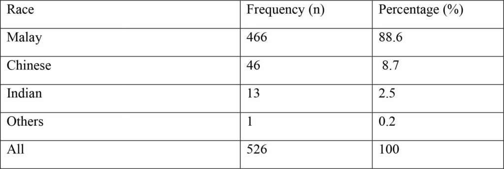
In present study total 526 CLP patients registered in the study hospital records were included. Majority (86.7%) of these patients were in the age group < 18years, most (56.7%) of them were females. The racial distribution of patients was 88.6 % Malays, 8.7 % Chinese, 2.5 % Indian and 0.2% others (Table. 1). Among Malay patients 77 % had CLP, 12 % had CP and 9 % had CL. In Chinese patients 76 % of cases were of CLP, 17 % CP and 6.5 % CL. Moreover, 76 % Indian had CLP as listed in Table 2. Furthermore, distribution of CP types among Malay, Chinese, Indian and other races is listed in Table 3. The occurrence of soft and hard palate cleft was more frequent affecting 92 % Malay,

Table 1: Race Distribution (n = 526)
Table 2: Distribution of facial cleft type among different races
Table 3: The distribution of cleft palate type according to races
Table 4: The distribution of cleft side in relation to different races

95 % Chinese and 69 % Indian study subjects. Furthermore, details about pattern of oral cleft side among study races is elucidated in Table 4. The bilateral oral cleft was found among 53.8 % Indians, 52.1% Chinese and 41.6 % Malay subjects.

DISCUSSION

Orofacial clefts significantly lead to long-term financial and psychological stress for affected subjects and their families. The surgical treatment for orofacial cleft involves multidisciplinary approach. The treatment begins shortly after birth and may continue up to teenage and comprise of multiple surgeries and long-term speech therapy combined with orthodontic treatment.11

The present study found that out of 526 CLP patients, the racial distribution of patients was 88.6 % Malays, 8.7 % Chinese, 2.5 % Indian and 0.2% others. Among Malay patients 77 % had CLP, 12 % had CP and 9 % had CL. In Chinese patients 76 % of cases were of CLP, 17 % CP and 6.5 % CL. Moreover, 76 % Indian had CLP. The results of present study are in agreement with findings previous studies who reported that CLP was most frequent type of oral cleft in Malaysian population.12,13 However, to best of authors’ knowledge no study was found in literature stating racial distribution of oral clefts in Malaysia. Moreover, these results of current study are consistent with several other epidemiologic studies.14-16 Furthermore, many other published studies report higher percentage of CLP compared to isolated CL and CP. The percentage of CLP was 66% in Brazil, 78.3% in Saudi Arabia, 76.8% in Sudan and in Mexico it was found to be 70%.17-20

The occurrence of soft and hard palate was more frequent affecting 92 % Malay, 95 % Chinese and 69 % Indian study subjects. The bilateral oral cleft was found among 53.8 % Indians, 52.1% Chinese and 41.6 % Malay subjects. In contrast to present study, unilateral cleft on left side was reported to be more predominant.21-23 Moreover, Aljohar et. al also reported occurrence of oral cleft on left-side more frequently, which is in agreement with a study conducted in China.24,25 In literature no any substantial justification for the frequent occurrence of left side cleft is found. However, it has been proposed that close proximity of blood vessels resulting in profuse blood supply to right side of the fetal head may be one reason for less occurrence of cleft on right side.26,27 It is recommended that molecular and genetic studies of Malaysian ethnic groups with oral clefts should be carried out to explore reasons for variations in prevalence of oral clefts among ethnic groups of Malaysia.

CONCLUSION

The right side oral cleft was found in 96.1% of Malay study subjects and 3.9 % Chinese. Among Chinese ethnic participants, majority (52 %) had bilateral oral cleft. Overall data indicates hard palate cleft was present in 3.7% Malay, 4.6% Chinese, and 23% Indian study participants. However, soft plate cleft was present in 4 % Malay and 7.6% Indian. Among ethnic groups 92% of Malay patients, 95% of Chinese and 69% of Indian had hard and soft palate cleft.

CONFLICT OF INTEREST

None declard

REFERENCES

  1. Conway JC, Taub PJ, Kling R, Oberoi K, Doucette J and Jabs EW, Ten-year experience of more than 35,000 orofacial clefts in Africa, BMC Paediatrics, 2015;15:8. https://doi.org/10.1186/s12887-015-0328-5
  2. Kirschner RE and Rossa, DL. Cleft lip and palate. Otolaryngol Clin North Am; 2000; 36 (6): 1191-1215. https://doi.org/10.1016/S0030-6665(05)70277-2
  3. Strong EB, Buckmiller LM. Management of the cleft palate. Facial Plast Surg Clin North Am; 2001; 9(1): 15-25.
  4. Stanier P, Moore GE. Genetics of cleft lip and palate: syndromic genes contribute to the incidence of non-syndromic clefts. Hum Mol Genet; 2004; 1: 73-81. https://doi.org/10.1093/hmg/ddh05
  5. World Health Organization, Global strategy to reduce the health-care burden of craniofacial anomalies. (Available on request from the Human Genetics Programme of the World Health Organization, 2002; 1211 Geneva 27, Switzerland.)
  6. Onyango JF, Noah S. Pattern of clefts of the lip and palate managed over a three-year period at a Nairobi hospital in Kenya. East Afr Med J, 2005; 82:649-651.
  7. Kozelj V, Vegnuti M. Time series analysis of births of children with orofacial clefts J Craniomaxillofac Surg, 2000; 28: 201-203. https://doi.org/10.1054/jcms.2000.0150
  8. Christensen K, Fogh-Andersen P, Etiological subgroups in non-syndromic isolated cleft palate. A genetic epidemiological study of 52 Danish birth cohorts. Clin Genet. 1994; 46(5):329-35. https://doi.org/10.1111/j.1399-0004.1994.tb04173.x
  9. Khoury MJ, Weinstein A, Panny S, Holtzman NA, Lindsay PK, Farrel K, Eisenberg M Maternal cigarette smoking and oral clefts: a population-based study. Am J Pub Health, 1987;77:623-625. https://doi.org/10.2105/AJPH.77.5.623
  10. Hammond M and Stassen L. Do you CARE? A national register for cleft lip and palate patients. Br J Plast Surg; 1999; 52 (1): 12-17. https://doi.org/10.1054/bjps.1998.3021
  11. Anderson S L, Adams G, Plaut VC, The cultural grounding of personal relationship: the importance of attractiveness in everyday life. J Person and Soc Psycho 2008;95:352 – 368. https://doi.org/10.1037/0022-3514.95.2.352
  12. Chai SC, Jimeno ZKL, Sasidaran R, Sergius A. Pilot epidemiological study of cleft lip and/or palate in Kota Kinabalu Sabah, Asian J of Medic Sci, 2013,4;86-91.
  13. Tan KB, Tan KH, Yeo GS. Cleft deformities in Singapore: a population-based series 1993-2002. Singapore Med J; 2008; 49(9): 710-714.
  14. Omo-Aghoja VW, Omo-Aghoja LO, et al. Antenatal determinants of oro-facial clefts in Southern Nigeria 2010 Mar;10(1):31-9.
  15. Elahi MM, Jackson IT, et al. Epidemiology of cleft lip and palate in Pakistan. Plast. Reconstr. Surg 2004;113(6):1548-1555.
    https://doi.org/10.1097/01.PRS.0000117184.77459.2B
  16. Yoshikazu Nagase, Nagato Natsume, et al. Epidemiological analysis of cleft lip and/or palate by cleft pattern. J. Maxillofac. Oral. Surg, 2010; 9(4):389-395. https://doi.org/10.1007/s12663-010-0132-6
  17. Barbosa D, Reis, Hercílio MJ, Letícia V, Portz Paulo R, Ferreti Bonan A. Prevalence of nonsyndromic oral clefts in a reference hospital in the state of Minas Gerais, Brazil, between 2000-2005. Braz Oral Res; 2007; 21 (4): 314-317. https://doi.org/10.1590/S1806-83242007000400006
  18. Al-Balkhi KM. The distribution and classification of clefts in patients attending a cleft lip and palate clinic in Riyadh, Saudi Arabia. Saudi Med J; 2008; 29 (5): 739-742.
  19. Suleiman AM, Hamzah ST, Abusalab MA and Samaan KT. Prevalence of cleft lip and palate in a hospital-based population in the Sudan. Int J Paediatr Dent; 2005;15(3):185- 189. https://doi.org/10.1111/j.1365-263X.2005.00626.x
  20. Blanca S, González M, L López M, A Rico, Fernando G. Oral clefts: a retrospective study of prevalence and predisposal factors in the state of Mexico. J of oral sci; 2008; 50 (2): 123 -129. https://doi.org/10.2334/josnusd.50.123
  21. Feliciona Blanco-Davila. Incidence of cleft lip and palate in Northeast of Mexico: A 10 years study. The J Craniofac Surg 2003;14(4):533-537. https://doi.org/10.1097/00001665-200307000-00027
  22.  Chuangsuwanich A, Aojanepong C, Muangsombut S,and Tongpiew P. Epidemiology of cleft lip and palate in Thailand. Ann Plast. Surg.1998;10:7.12.
  23. Ogle, O.E. Incidence of cleft lip and palate in a newborn Zairian sample. Cleft palate Craniofac J. 1993;30:250.
  24.  Srivastava S., and Bang, R.L. Facial clefting in Kuwait and England: A comparative study. Br. J. Plast. Surg. 1990;43:457. https://doi.org/10.1016/0007-1226(90)90013-P
  25. Aljohar A, Ravichandran K, Subhani S, Pattern of Cleft Lip and Palate in Hospital-Based Population in Saudi Arabia: Retrospective Study, Cleft Palate-Craniofacial J, 2008, 45; 6:592-6. https://doi.org/10.1597/06-246.1
  26. Zhou QJ, Shi B, Shi ZD, Zheng Q, Wang Y. Survey of the patients with cleft lip and palate in China who were funded for surgery by the Smile Train program from 2000 to 2002. Chin Med J. 2006; 119:1695-1700.
  27.  Johnston C, Brown KS. Human population data. General discussion III. Prog Clin Biol Res 1980; 46.117 131.

1. Associate Professor, Department of Oral Medicine and Periodontology, Bibi Aseefa Dental College, SMBB Medical University Larkana.
2. Assistant Professor and Head Department of Science of Dental Materials, Bibi Aseefa Dental College, SMBB Medical University Larkana.
3. Assistant Professor, Community Dentistry, Bibi Aseefa Dental College, SMBB Medical University Larkana.
Corresponding author: “Dr. Shahid Ali Mirani ” <shahid.ali@lumhs.edu.pk> </shahid.ali@lumhs.edu.pk>

Evaluating Occurrence of Variable Cleft Lip and Palate Types Among Ethnic Groups of Malaysia

Syed Yousif Ali Shah                   BDS, MDSc

Shahid Ali Mirani                        BDS, MPhil, PhD

Muhammad Amin Sahito           BDS, MSc

OBJECTIVE:

The objective was to assess the distribution of types of cleft lip and palate (CLP) among different racial groups living in Malaysia.

METHODOLOGY: This study was carried out in two tertiary care hospitals of Malaysia, during August 2007 to March 2009. Total 526 CLP patients registered in the hospital records during the study period were included. The Modified Craniofacial Anomalies Registration (CARE) form was used to collect data of different types of oral clefts in relation to race among Malaysian.

RESULTS: Of the total 526 patients registered in the study hospital records during the study period. Majority (86.7%) of these patients were in the age group < 18years, most (56.7%) of them were females. The racial distribution of patients was 88.6 % Malays, 8.7 % Chinese, 2.5 % Indian and 0.2% others. The right side oral cleft was found in 96.1% of Malay study subjects and 3.9 % Chinese. Among Chinese ethnic participants, majority (52 %) had bilateral oral cleft. Overall data indicates hard palate cleft was present in 3.7% Malay, 4.6% Chinese, and 23% Indian study participants. However, soft plate cleft was present in 4 % Malay and 7.6% Indian. Among ethnic groups 92% of Malay patients, 95% of Chinese and 69% of Indian had hard and soft palate cleft.

CONCLUSION: Ceft lip, cleft palate, race, ethnic, Malaysia

KEY WORDS:White spot lesion, Fluoride bonding agent RMGICs, Casein phosphopeptide amorphous calcium phosphate, Resin infiltration.

HOW TO CITE: Shah SYA, Mirani SA, Sahito MA. Evaluating occurrence of variable cleft lip and palate types among ethnic groups of malaysia. J Pak Dent Assoc 2018;27(1):9-12.

DOI: https://doi.org/10.25301/JPDA.271.9
Received:
23 November, 2017, Accepted: 29 December, 2017

Managing White Spot Lesion During and After The Orthodontic Treatment

Fatama Siddika                                                      PHD

Mohammad Sayedur Rahman Khan                    BDS

Ren Jia Bao                                                            BDS

Ma Wen Sheng                                                      BDS

ABSTRACT:

The constitution of enamel demineralization or white spot lesion (WSL) at the side of fixed orthodontic appliance is a usual complexity seen at the period of fixed orthodontic treatment and after completion of this treatment, which scupper the accomplishment of a successfully completed case. Restraining programmes for managing this orthodontic side effect must be accentuated for all orthodontic patients. The concern of an orthodontist is to abate the risk of decalcification as a sequence of orthodontic treatment by instructing and motivating the patients for good oral hygiene care. Recognition of WSL before the orthodontic treatment might allow performing preventive measures to counteract the process of progression of lesion. Modern strategies of managing WSL during and after the orthodontic treatment are reviewed coevally in this article. This review represents the prevention and management of white spot lesion by topical fluoride releasing materials (bonding agent resin modified glass ionomer cements [RMGICs], toothpaste, gel, mouthwash, varnishes), pro-biotic, polyols, antiseptic, sealants, casein supplements, laser, tooth bleaching agent, micro-abrasion and resin infiltration which are effectual. Nevertheless the implementation of RMGICs, casein phosphopeptide amorphous calcium phosphate, micro-abrasion, resin infiltration can be more adjuvant than only the fluoride rinses in the abatement
of white spot lesion or demineralization spots.

KEY WORDS:White spot lesion, Fluoride bonding agent RMGICs, Casein phosphopeptide amorphous calcium phosphate, Resin infiltration.

HOW TO CITE: Siddika F, Khan MSR, Bao RJ, Sheng MW. Managing white spot lesion during and after the orthodontic treatment. J Pak Dent Assoc 2018;27(1):1-8.

DOI: https://doi.org/10.25301/JPDA.271.1

Received: 29 October, 2017, Accepted: 29 December, 2017

INTRODUCTION

Orthodontic fixed appliances and enamel demineralization have a strong relation with each other in orthodontic treatment. Enamel demineralization is the initial exposure of carious lesion which can be identified with naked eye as white spot lesion (WSL).1
In most cases it located at the side of the bands and brackets (Figure-1, Figure-2). This initial lesion can extend to cavitated lesion from enamel to dentin. Diagnosis of this lesion can be done by Meticulous visual examination, the white intensity as well as limit and opacity features under transillumination. On the basis of diagnosis this lesion can be classified into following types: superficial(non-cavitated), mixed(non-cavitated and cavitated ) and deep (cavitated).

These lesions are non fluoridated opacities with defined shape and well differentiated from surrounding enamel. It is usually seen in the orthodontic treatment due to improper
oral hygiene. Oral hygiene is impeded by orthodontic appliances (e.g. bands, brackets, wires) which create retention areas for dental plaque because of their irregular surfaces as well as making chance to elongate the retention of plaque on surfaces of teeth.2-6 This elongation of plaque retention along with acidogenic bacteria such as streptococcus mutans, and various lactobacilli play a momentous role in the development of enamel demineralization or white spot lesion.7-8 Previous clinical study showed WSLs occurrence highly increase in first 6 months of treatment and continuing slowly up to 12 months, for this reason appropriate maintenance of oral hygiene is consulted at the starting of the orthodontic treatment.9

Other factors associated with higher risk of WSLs are younger age, prolong treatment duration, percentage of treatment time in elastomeric chain, number of missed appointments, patients with malocclusion, number of poor oral hygiene excerpt in the patient chart, poor attending, male gender and poor oral hygiene at the screening examination.10 The aesthetic purpose of treatment is hampered by this white spot lesion.6,11-13 Improving facial and dental aesthetic is the first priority of all Orthodontist which can mar with the appearance of white spot lesion after removal of orthodontic appliances. Hence Orthodontist and patient ought to treat to impede the formation of white spot lesions. Several techniques for the management of WSLs during and after the orthodontic treatment are implemented, but the foremost one is still controversial.

This side effect of orthodontic treatment affects patient’s aesthetic as well as smile and can be very difficult or sometimes even impossible to eradicate after detaching fixed appliances. After completion of orthodontic treatment, the appearance of WSLs is about 50% and the aim of all Orthodontist is to annihilate WSLs.14-20 Prevalence of WSLs usually associated with the labio-buccal surfaces of maxillary teeth with this manner – lateral incisors, canines, premolars and central incisors.21,22 Earlier study showed that WSLs occur both on labial and lingual surfaces of teeth.23 WSLs mostly found on maxillary anterior segment and very few on maxillary posterior segments, in addition females are less affected then male.24 Incidence and prevalence of WSLs between right and left sides of the maxilla and mandible do not have any special difference.25-27 The aim of this study is to oversee the ways of managing WSLs based on the current information during and after the orthodontic treatment.

PATHOGENESIS OF WSLS

The term WSL was defined as “Caries like lesion which can be seen on enamel by naked eye as first sign”.28 Remarkable alteration usually happen in concentration of acidogenic bacteria (especially Streptococcus mutans and lactobacilli) while orthodontic appliances are applied into the oral cavity. Orthodontic patient with plaque of higher concentration of acidogenic bacteria has greater extent in comparison with other patients which causes progressive caries.29

ETIOLOGY

The prevalence of WSL on the enamel surface is caused by multiple factors in the period of fixed orthodontic treatment. Presence of bacterial plaque, fermentable carbohydrates, a susceptible tooth surface and a sufficient period of time are necessary for the formation of WSL. Many irregularities are present in the fixed appliance of orthodontic treatment which causes easy accumulation of food debris along with the cariogenic bacteria and cause the manual teeth cleansing more difficult as well as also avert the selfcleansing action of tongue, lips and cheeks to remove accumulated food debris from the tooth surfaces. The gingival side of brackets are the area of great plaque deposition.30 Elastomeric rings ligated teeth have greater number of cariogenic microorganism arrangements which can be seen fewer in stainless steel ligature wires ligated teeth.31

PREVENTION AND MANAGEMENT OF WSLS

The prevention and management of white spot lesions can be achieved by applying multifunctional approaches during orthodontic treatment. Mechanical plaque control methods for improving patient oral hygiene The most important prophylactic measure is maintenance of good oral hygiene to prevent the occurrence of WSLs in orthodontic patient .The commencement of prevention can be achieved by educating and motivating the patient to noncariogenic diet as well as by observing of oral hygiene for compliance. Tooth brushing of tooth surfaces daily two times at least, with fluoride containing toothpaste is highly advised for mechanical Plaque controlling and removing the plaque from retaining areas. For attaining patient good oral hygiene a standard modified tooth brush, uses of disclosing solutions and floss can aid. More effective method of reduction of plaque accumulation might be daily water irrigation or use of a power toothbrush in combination with manual tooth brushing than manual tooth brushing only.32 To enhance the brushing efficacy and facilitating the patient cleaning as well as to reduce the bacterial load, professional prophylactic cleaning is quite effective. Professional tooth cleaning two or three times yearly can abate the risk of caries in number of teeth and keep up a healthy mouth. The inaccessible areas which are difficult to brush for the patient can be properly cleaned by professional tooth cleaning. Polishing of the coronal surfaces by progressively finer particle size fluoridated pastes and brushes or polishing elastomer cups can prevent
the bacterial mechanical retention.33

Enamel resistance can be increased against the microbial acids by applying topical fluoride

Complacent action of fluoride in resisting WSLs has been proved with the uses of the followings such as fluoride mouthwashes, fluoride gel, fluoride toothpaste, fluoride varnishes, fluoride in bonding agents and fluoride in elastomers. White spot lesions/dental caries can be impeded at early stages by fluoride ions through several mechanisms like alteration of bacterial metabolism of dental plaque through inhibition of some enzymatic processes, inhibition of production of acids, alteration of the composition of bacterial flora and/or the metabolic activity of microorganisms; and decreasing demineralization as well as increasing re-mineralization of white spot lesion.34

Fluoride containing bonding agent

Risk of caries increases in case of prolong orthodontic treatment patient. If the bonding system incessantly releases fluoride at the side of bracket base, it would be immensely adjuvant. Resin modified glass ionomer cements (RMGICs) have been proposed as bracket bonding agents due to their continuous fluoride releasing properties throughout the orthodontic treatment. RMGICs continuously take up fluoride from the environment (e.g. fluoride in dentifrices, in oral rinse and in potable fluoridated water), act as pumps and precisely re-release fluoride to the most prone areas of WSLs. The application of RMGICs is performed in the following manner – first enamel de-proteinization (to remove the surface organic layer) is done by application of 5.25% sodium hypochlorite (NaOCL) on enamel surface for 1 min and then etched with 37% phosphoric acid for 30s, this creates type 1 etching pattern (the enamel rod, or prism, heads are dissolved) and type 2 etching pattern (in which the enamel inter-prismatic substance is dissolved) forms micro-porosities of increase number which allow the bonding agent (RMGICs) to penetrate the enamel surface for enhancing the bracket bonding rate of success (by increasing shear bond strength [SBS]) as well as serve the added benefit of minimizing WSL development.

Fluoride containing Toothpaste

Fluoride toothpaste containing either one of these (sodium fluoride, monoflourophosphate, stannous fluoride) or a combination of these compounds is recommended. In comparison with normal formulation (more than 1000 ppm) the 5000 ppm concentration of fluoride in toothpaste is more effective to treat WSL.35-37 Due to the application of fluoride ion on enamel surface a fluoroapatite crystal structure is formed which is less soluble with hydroxyapatite in the oral environment. Use of fluoride toothpaste is mostly recommended regularly to the patients by the orthodontist as they are at high risk of WSLs, only fluoride toothpaste uses is not effective in resisting WSL in the majority of patients, even with good oral hygiene implementation of other sources fluoride together with fluoride tooth paste is recommended.5,38-43

Flouride containing mouthwashes

Everyday uses of fluoridated mouthwash (contain 0.05% sodium fluoride) caused significant abatement of the lesion formation at the side of brackets and beneath the bands. Nowadays antimicrobial agents like chlorhexidine, zinc, triclosan conjoin with mouthwashes to enhance cariostatic effects. Previous study recommended, prevention of enamel demineralization during fixed orthodontic treatment by the everyday used of 0.05% NaF mouthwash has the best strategy,44 also everyday rinsing mouth with NaF 0.05% or 0.2% and/or weekly rinse with AFP(1.2%)have been observed reducing WSLs incidence during active fixed orthodontic treatment.45 Fluoride concentration in the saliva significantly increased if NaF mouthwash uses for 2 week (one rinse per day).44

Fluoride containing gel

Use of 1100ppm fluoride toothpaste together with 0.4% stannous fluoride gel applied twice daily by toothbrush, both provide additional protection against decalcification.46

Fluoride containing pits and fissure sealants

Placing of light cured pit and fissure sealants adjacent to bonded orthodontic brackets on the labial/buccal surface were 80% effective in resisting white spot lesion was found in vitro and required no patient compliance.47 It is quite difficult to remove the sealant and after removal it needs precision polishing.

Fluoride varnish

The efficacy of fluoride varnishes have proved largely in decreasing the incidence of caries. In poorly-motivated patients, this varnish can be applied with an intensive treatment schedule (every three to four monthly). Remission of demineralization of the enamel at the side of brackets, enhancing re-mineralization of the carious lesions as well as impediment of further lesions can be achieved by the fluoride varnishes. Inspite of patient’s noncompliance, topical fluoride varnish is quite advantageous for preventing WSLs and delivering the fluoride in a sustained manner over a longer period of time. Biannually Duraphate applying to WSLs areas showed 30-50% reduction in comparison with the non-varnished areas.48 Application of fluoride varnish  with excellent oral hygiene to prevent WSLs has been proved as fast and simple technique.

Additional method’s different mechanisms

Probiotics

Pro-biotics are live microorganisms with health benefits when they are administered in adequate numbers inhibit other microorganisms, especially pathogens. Probiotic bacteria might enhance effect of fluoride in preventing dental caries.49

Polyols

Polyols are weakly metabolized (sorbitol) or nonmetabolized (xylitol) by cariogenic bacteria. Chewing gum with xylitol (2 g of xylitol/socket) or polyols is recommended after each meal (three times daily) for 10-20 min.45-52 Previous study reported that xylitol lozenges significantly decreased the acidity of dental plaque in fixed orthodontic appliance patients after consumption of sucrose.50

Antiseptic (Chlorohexidine varnish)

Chlorohexidine is available in different presentation like mouthwashes, gel or varnishes. Chlorohexidine varnishes are more effective than its gels and mouthwashes, chlorohexidine varnish reduces streptococcus mutans levels in orthodontic patients with fixed appliances and therefore might be useful to prevent carious lesion.53When combining chlorohexidine varnish with a fluoride varnish (Fluor Protector), the cariostatic effect becomes enhanced even in comparison to ozone, which also can decrease streptococcus mutans count.54

Use of casein phosphopeptides amorphous calcium phosphate

Recent research has shown that casein phosphopeptidesamorphous calcium phosphate (CPP-ACP) absorb through the enamel surface and affected the demineralization-remineralization processes,55 this processes is performed by CPP which is derived from casein protein and contains calcium and phosphate ions “stuck” to create CPP-ACP. The calcium and phosphate ions are provided by this CPP-ACP and shows anti-cariogenic activity on tooth surface, thereabout CPP-ACP knows as store house of calcium and phosphate.53 The calcium and phosphate ions are released while the occurrence of intraoral acid attack and aid the saliva to obtain a supersaturated state of ions which precipitate a calciumphosphate compound on the exposed tooth surface.56 In addition, ammonia is produced by the disintegration of the CPP lead to increase the pH (buffer) and delays formation of bio-film by preventing bacterial adhesion to tooth surfaces.57 In CPP-ACP mechanism, deeper penetration of ions results the entire body re-mineralization of the lesion inspite of acting only on the surface layer, thus the aesthetic appearance improves. A recent study showed that application of CPP-ACP-containing varnish to incisors, with or without brushing and use of a mouthwash, decreased the depth of carious lesions around orthodontic brackets. 5 8

Lasers

Laser beams increase enamel micro-hardness and resistance to acid attack. In preventive dentistry the argon lasers, CO2, Nd-YAG, and erbium YAG are used.50,59,60 Irradiation of enamel decreases amount of demineralization up to 30%-50% with argon laser beams,61 laser beams also lowered the dissolution threshold pH value. Laser beams maintain an intact enamel surface but cause surface morphology changes. Exposure to laser beams micro-spaces form within the enamel, the released ions trap into these micro-spaces and act as re-mineralization sites within the enamel surface. The mean lesion depth is significantly reduced by the application of argon laser beams (488 nm) compared to visible light controls.62

AFTER ORTHODONTIC TREATMENT

After the removal of fixed orthodontic appliances, a regressive appearance of WSLs might be seen because of salivary radical re-mineralization as well as brushing abrasion and such progress relies on the severity of lesions.63 Nevertheless this WSLs re-mineralization is not sufficient to overcome the lesion without definitive treatment.

Re-mineralization

Re-mineralization is the initial step of WSLs improvement. Several products are available for such purpose in different forms like solutions, varnishes, creams, pastes and chewing gums which contain fluorides and/or casein phosphopeptideamorphous calcium phosphate and are necessary to apply more invasive techniques such as resin-infiltration, bleaching and micro-abrasion. This lesion should not be treated by the products containing higher concentrations of fluoride in incisors and canines as those caused tooth discoloration.64

Bleaching

Bleaching procedures show short range of aesthetic outcome and cause less enamel micro-hardness as well as teeth sensitivity.65 Nevertheless bleaching with 10% carbamide peroxide as well as adding casein phosphopeptide-amorphous calcium phosphate is considered to incipient enamel caries without having any effect on enamel properties (mechanical and chemical) along with promoting in mineral gaining on subsurface of lesion.66 Bleaching with bleaching agents along with three different types of biomaterials such as nanohydroxyapatite, nano-BAG and nano-amorphous calcium phosphate soothe the unsatisfactory effects of bleaching as well as impede the irreversible damage over the enamel surface.67,68

Micro-abrasion

Micro-abrasion alone with the bleaching technique is an active method for the treatment of post orthodontic WSLs while the depth of lesion is less than 0.2 mm.69 After debonding, micro-abrasion plays a good role by the process of salivary re-mineralization and continuous surface abrasion for improving lesions because of its invasive nature. While applying abrasive slurry of 6.6% (Opalustre) or 6% (Whiteness RM) hydrochloric acid with a brush on the enamel surface, it shows chemical and mechanical effects.70

Resin-infiltration

A low-viscosity resin is used to infiltrate the WSL after completion of several steps like etching with 15% HCl acid for 20s then rinsing, drying and dehydration of the enamel surface with ethanol; which makes the enamel surface more permeable for triethylene glycol dimethacrylate-based resin to pass into the underlying porous structure caused to mask the WSL as well as reinforcing prismic structure of enamel.69 The masking effects rely on the depth of the lesion and cosmetic outcome of the treatment is more satisfactory in the beginning level known as active stage.71,72 1 year followup study has shown that permanent cosmetic improvement can be achieved in post-orthodontic WSLs with this method.73
Other study has seen that no colour change occured after 2, 6, and 12 months, consecutively.74 However, previous study showed that satisfactory clinical performance can be obtained after 19 months from the treatment of WSLs associated with non-orthodontic etiology such as fluorosis, after trauma.75 An earlier study evaluated the colour improvement and stability of WSLs treated by fluoride, micro-abrasion or infiltration and found the acceptable result in favour of infiltration and micro-abrasion.70

CONCLUSION

The common side effect of fixed orthodontic treatment is WSLs. Therefore the responsibility of an orthodontist is to abate the risk of the patient having decalcification as a consequence of orthodontic treatment by instructing and motivating the patients for practicing excellent oral hygiene. Prophylaxis and management should be carried out by using high-fluoride toothpaste, gel, mouthwash, varnishes application, bonding agent RMGICs, pro-biotic, polyols, antiseptic, sealants, casein supplements, laser, tooth bleaching, micro-abrasion and resin infiltration.

Disclosure of funding: Nil

Conflict of Interest: Nil

REFERENCES

  1.  Fejerskov O, Nyvad B, Kidd EAM. Clinical and histological manifestations of dental caries. In Fejerskov O, Kidd EAM, editors. Dental Caries. The disease and its clinical management. Copenhagen: Blackwell Munksgaard. 2003; pp. 71-99
  2. Batoni G,Pardini M,Giannotti A, Ota F, Guica MR, Gabrielle M.Effect of removable orthodontic appliances on oral colonisation by mutansstreptococciinchildren.Eur J Oral Sci 2001;109:388-92. https://doi.org/10.1034/j.1600-0722.2001.00089.x
  3. Petti S,Barbato E, Simonettid’ArcaA.Effect of orthodontic therapy with fixed and removable appliances on oral microbiota: a six-month longitudinal study.New Microbial 1997;20;55-62.
  4. Rosenbloom RG, Tinanoff N, Salivary streptococcus mutans levels in patients before,during,and after orthodontic treatment. Am J OrthodDentofacialOrthop 1991;100:35-7. https://doi.org/10.1016/0889-5406(91)70046-Y
  5. Maxfield BJ, HamdanAM, Tüfekçi E, Shroff B, Best AM, Lindauer SJ. Development of white spot lesions during orthodontic treatment: perceptions of patients, parents, orthodontists, and general dentists. Am J OrthodDentofacialOrthop. 2012 Mar; 141(3): 337-44
  6. Lucchese A, Gherlone E. Prevalence of white-spot lesions before and during orthodontic treatment with fixed appliances. Eur J Orthod. 2013 Oct;35(5):664-8. https://doi.org/10.1093/ejo/cjs070
  7. Srivastava K, Tikku T, Khanna R, Sachan K. Risk factors and management of white spot lesion in orthodontics. J OrthodSci 2013;2:43-49. https://doi.org/10.4103/2278-0203.115081
  8. ØggardB.Prevalance of white spot lesions in 19-yearsolds;a study on untreated and orthodontically treated persons 5 years after treatment. Am J OrthodDentofacialOrthop 1989;96:423-7. https://doi.org/10.1016/0889-5406(89)90327-2
  9. Tufekci E, Dixon JS, Gunsolley JC, Lindauer SJ. Prevalence of white spot lesions during orthodontic treatment with fixed appliances. Angle Orthod. 2011; 81: 206-10. https://doi.org/10.2319/051710-262.1
  10. Chapman JA, Roberts WE, Eckert GJ, Kula KS, Gonzàlez-Cabezas C. Risk factors for incidence and severity of white spot lesions during treatment with fixed orthodontic appliances. Am J OrthodDentofacialOrthop 2010;138:188- 94. https://doi.org/10.1016/j.ajodo.2008.10.019
  11. Arjun J, Brobakken BO, Prevalence of carious white spots after orthodontic treatment with multibondedappliances.Eur J Orthod 1986;8:229-34.
    https://doi.org/10.1093/ejo/8.4.229
  12. O’Reilly MM,Featherstone JDB. Decalcification and remineralization around orthodontic appliances:an in vivo study.Am J OrthodDentofacialOrthop 1987;92:33-40. https://doi.org/10.1016/0889-5406(87)90293-9
  13. ShunginD,OlssonAI,Perrson M Orthodontic treatmentrelated white spot lesions:a 14-year prospective quantitative follow-up, including bonding material assessment.Am J OrthodDentofacialOrthop 2010;138:136.el-8.
  14. Karlinsey RL, Mackey AC, Stookey GK, Pfarrer AM. In vitro assessments of experimental NaF dentifrices containing a prospective calcium phosphate technology. Am J Dent. 2009 Jun;22(3):180-4.
  15. Summit JB, Robbins JW, Schwartz RS. 3rd ed.ch.1. Hanover Park, IL: Quintessence publishing;2006. Fundamentals of Operative Dentistry:A Contemporary Approach;pp.2-4.
  16. Øgaard B, Bishara SE, Duschner H. Enamel effects during bonding-debonding and treatment with fixed appliances. In: Graber TM, Eliades T, Athanasiou AE: Risk Management in Orthodontics: Experts Guide to Malpractice. Hanover Park, IL, Quintessence. 2004; 19-46
  17. Lundström F, Krasse B. Streptococcus mutans and lactobacilli frequency in orthodontic patients; the effect of chlorhexidine treatments. Eur J Orthod. 1987;9:109-16. https://doi.org/10.1093/ejo/9.2.109
  18. Scheie AA, Arneberg P, Krogstad O. Effect of orthodontic treatment on prevalence of Streptococcus mutans in plaque and saliva. Scand J Dent Res. 1984;92:211-7. https://doi.org/10.1111/j.1600-0722.1984.tb00881.x
  19.  Zachrisson BU, Zachrisson S. Caries incidence and oral hygiene during orthodontic treatment. Scand J Dent Res. 1971;79:394-401.
    https://doi.org/10.1111/j.1600-0722.1971.tb02028.x
  20. Sakamaki ST, Bahn AN. Effect of orthodontic banding on localized oral lactobacilli. J Dent Res. 1968;47:275-9. https://doi.org/10.1177/00220345680470021301
  21. Balenseifen JW, Madonia JV. Study of dental plaque in orthodontic patients. J Dent Res. 1970;49:320-4. https://doi.org/10.1177/00220345700490022101
  22. Lundström F, Krasse B. Caries incidence in orthodontic patients with high levels of Streptococcus mutans. Eur J Orthod. 1987;9:117-21.
    https://doi.org/10.1093/ejo/9.2.117
  23. Chang HS, Walsh LJ, Freer TJ. The effect of orthodontic treatment on salivary flow, pH, buffer capacity, and levels of mutans streptococci and lactobacilli. AustOrthod J. 1999;15:229-34.
  24. Türkkahraman H, Sayin MO, Bozkurt FY, Yetkin Z, Kaya S, Onal S. Archwire ligation techniques, microbial colonization, and periodontal status in orthodontically treated patients. Angle Orthod. 2005;75:231-6.
  25. Sukontapatipark W, el-Agroudi MA, Selliseth NJ, Thunold K, Selvig KA. Bacterial colonization associated with fixed orthodontic appliances. A scanning electron microscopy study. Eur J Orthod. 2001;23:475-84. https://doi.org/10.1093/ejo/23.5.475
  26. Smales RJ. Plaque growth on dental restorative materials. J Dent. 1981;9:133-40. https://doi.org/10.1016/0300-5712(81)90087-7
  27. Gwinnett AJ, Ceen RF. Plaque distribution on bonded brackets: A scanning microscope study. Am J Orthod. 1979;75:667-77. https://doi.org/10.1016/0002-9416(79)90098-8
  28. Forsberg CM, Brattström V, Malmberg E, Nord CE. Ligature wires and elastomeric rings: Two methods of ligation, and their association with microbial colonization of Streptococcus mutans and Lactobacilli. Eur J Orthod. 1991;13:416-20. Streptococcus mutans and Lactobacilli. Eur J Orthod.
    1991;13:416-20. https://doi.org/10.1093/ejo/13.5.416
  29. Lundström F, Krasse B. Streptococcus mutans and lactobacilli frequency in orthodontic patients; the effect of chlorhexidine treatments. Eur J Orthod. 1987;9:109-16. https://doi.org/10.1093/ejo/9.2.109
  30. Gwinnett AJ, Ceen RF. Plaque distribution on bonded brackets: A scanning microscope study. Am J Orthod.
    1979;75:667-77. https://doi.org/10.1016/0002-9416(79)90098-8
  31. Forsberg CM, Brattström V, Malmberg E, Nord CE. Ligature wires and elastomeric rings: Two methods of ligation, and their association with microbial colonization of Streptococcus mutans and Lactobacilli. Eur J Orthod. 1991;13:416-20. https://doi.org/10.1093/ejo/13.5.416
  32. Harvey WJ, Powell KR. Care of dental enamel for the orthodontic patient. AustOrthod J. 1981; 7: 70-6. 16.
  33. Øgaard B, Rolla G, Øgaard B. Oral microbiological changes, long-term enamel alterations due to decalcification, and caries prophylactic aspects, in Brantley WA, Eliades T, eds: Orthodontic Materials: Scientific and Clinical Aspects. Stuttgart, Thieme. 2001; 123-142.
  34. Davidson CL, Bekke-Hoekstra IS. The resistance of superficially sealed enamel to wear and carious attack in vitro. J Oral Rehabil. 1980;7:299-305. https://doi.org/10.1111/j.1365-2842.1980.tb00448.x
  35. Al-Mulla A, Karlsson L, Kharsa S, Kjellberg H, Birkhed D. Combination of high-fluoride toothpaste and no ostbrushing water rinsing on enamel demineralization using an in-situ caries model with orthodontic bands. ActaOdontol Scand. 2010;68:323-8. https://doi.org/10.3109/00016357.2010.512863
  36. Bergstrand F, Twetman S. A review on prevention and treatment of post-orthodontic white spot lesions Evidencebased methods and emerging technologies. Open Dent J. 2011;5:158-62. https://doi.org/10.2174/1874210601105010158
  37. Feng CH, Chu XY. Efficacy of one year treatment of icon infiltration resin on post-orthodontic white spots. Beijing Da XueXueBao. 2013;45:40-3.
  38. Richter AE, Arruda AO, Peters MC, Sohn W. Incidence of caries lesions among patients treated with comprehensive orthodontics. Am J OrthodDentofacialOrthop. 2011;139:657- 64. https://doi.org/10.1016/j.ajodo.2009.06.037
  39. Bishara SE, Ostby AW. White spot lesions: Formation, prevention, and treatment. SeminOrthod. 2008;14:174-82. https://doi.org/10.1053/j.sodo.2008.03.002
  40. Sudjalim TR, Woods MG, Manton DJ. Prevention of white spot lesions in orthodontic practice: A contemporary review. Aust Dent J. 2006;51:284-9. https://doi.org/10.1111/j.1834-7819.2006.tb00445.x
  41. Bahoum A, Bahije L, Zaoui F. Enamel demineralization in orthodontics. Systematic use of fluoride in prevention and treatment. SchweizMonatsschrZahnmed. 2012;122:937-47.
  42. Benson PE, Parkin N, Dyer F, Millett DT, Furness S, Germain P. Fluorides for the prevention of early tooth decay (demineralised white lesions) during fixed brace treatment. Cochrane Database Syst Rev. 2013;12:CD003809. https://doi.org/10.1002/14651858.CD003809.pub3
  43. Benson PE, Parkin N, Millett DT, Dyer FE, Vine S, Shah A. Fluorides for the prevention of white spots on teeth during fixed brace treatment. Cochrane Database Syst Rev. 2004;3:CD003809. https://doi.org/10.1002/14651858.CD003809.pub2
  44. Chadwick BL, Roy J, Knox J, Treasure ET. The effect of topical fluorides on decalfication in patients with fixed orthodontic appliances: a systematic review. Am J OrthodDentofacialOrthop. 2005; 128: 601-6. https://doi.org/10.1016/j.ajodo.2004.07.049
  45. Gwinnett AJ, Buonocore MG, Sheykoleslam Z. Effect of fluoride on etched human and bovine tooth enamel surfaces as demonstrated by scanning electron microscopy. Arch Oral Biology. 1972; 17: 271-8. https://doi.org/10.1016/0003-9969(72)90210-5
  46. Boyd RL. Comparison of three self-applied topical fluoride preparations for control of decalcification. Angle Orthod.1993;63:25-30. .
  47. Garcia-Godoy F. Shear bond strength of a resin composite to enamel treated with an APF gel. Pediatric entistry. 1993; 15(4): 272-4.
  48. Demito CF, Rodrigues GV, Ramos AL, Bowman SJ. Efficacy of a fluoride varnish in preventing whitespot lesions as measured with laser fluorescence. J ClinOrthod. 2011; 45: 25-9.
  49. Twetman S, Keller MK. Probiotics for caries prevention and control. Adv Dent Res. 2012;24:98-102. https://doi.org/10.1177/0022034512449465
  50. Sengun A, Sari Z, Ramoglu SI, Malkoç S, Duran I. Evaluation of the dental plaque pH recovery effect of a xylitol lozenge on patients with fixed orthodontic appliances. Angle Orthod. 2004;74:240-4.
  51. Beyth N, Redlich M, Harari D, Friedman M, Steinberg D. Effect of sustained-release chlorhexidine varnish on Streptococcus mutans and Actinomycesviscosus in orthodontic patients. Am J OrthodDentofacialOrthop. 2003;123:345-8. https://doi.org/10.1067/mod.2003.19
  52. Kronenberg O, Lussi A, Ruf S. Preventive effect of ozone on the development of white spot lesions during multibracket appliance therapy. Angle Orthod. 2009;79:64-9. https://doi.org/10.2319/100107-468.1
  53. Shen P, Cai F, Nowicki A, Vincent J, Reynolds EC. Remineralization of enamel subsurface lesions by sugar-free chewing gum containing casein phosphopeptide-amorphous calcium phosphate. J Dent Res. 2001;80:2066-70. https://doi.org/10.1177/00220345010800120801.
  54. Cross K, Huq N, Stanton D, Sum M, Reynolds E. NMR studies of a novel calcium, phosphate and fluoride delivery vehicle-? S1-casein (59-79) by stabilized amorphous calcium fluoride phosphate nanocomplexes. Biomaterials. 2004;25:5061-9. https://doi.org/10.1016/j.biomaterials.2004.01.045
  55. Reynolds EC. Casein phosphopeptide-amorphous calcium phosphate: The scientific evidence. Adv Dent Res. 2009;21:25-9. https://doi.org/10.1177/0895937409335619
  56. Iijima Y, Cai F, Shen P, Walker G, Reynolds C, Reynolds EC. Acid resistance of enamel subsurface lesions remineralized by a sugar-free chewing gum containing casein phosphopeptide-amorphous calcium phosphate. Caries Res. 2004;38:551-6. https://doi.org/10.1159/000080585
  57. Cochrane NJ, Cai F, Huq NL, Burrow MF, Reynolds EC. New approaches to enhanced remineralization of tooth
    enamel. J Dent Res. 2010;89:1187-97. https://doi.org/10.1177/0022034510376046
  58. Pithon MM, Santos Mde J, de Souza CA, LeãoFilho JC, Braz AK, de Araujo RE, et al. Effectiveness of fluoride sealant in the prevention of carious lesions around orthodontic brackets: An OCT evaluation. Dental Press J Orthod. 2015;20:37-42. https://doi.org/10.1590/2177-6709.20.6.037-042.oar
  59. Olivi G, Genovese MD, Caprioglio C. Evidence-based dentistry on laser paediatric dentistry: Review and outlook. Eur J Paediatr Dent. 2009;10:29-40.
  60. Verma SK, Maheshwari S, Singh RK, Chaudhari PK. Laser in dentistry: An innovative tool in modern dental practice. Natl J Maxillofac Surg. 2012;3:124-32. https://doi.org/10.4103/0975-5950.111342
  61. Fox JL, Yu D, Otsuka M, Higuchi WI, Wong J, Powell GL. Initial dissolution rate studies on dental enamel after CO2 laser irradiation. J Dent Res. 1992;71:1389-98. https://doi.org/10.1177/00220345920710070701
  62. Noel L, Rebellato J, Sheats RD. The effect of argon laser irradiation on demineralization resistance of human enamel adjacent to orthodontic brackets: An in vitro study. Angle Orthod. 2003;73:249-58.
  63. Guzmán-Armstrong S, Chalmers J, Warren JJ. Ask us. White spot lesions: Prevention and treatment. Am J OrthodDentofacialOrthop. 2010;138:690-6. https://doi.org/10.1016/j.ajodo.2010.07.007
  64. Morrier JJ. White spot lesions and orthodontic treatment. Prevention and treatment. Orthod Fr. 2014;85:235-44. https://doi.org/10.1051/orthodfr/2014016
  65. Senestraro SV, Crowe JJ, Wang M, Vo A, Huang G, Ferracane J, et al. Minimally invasive resin infiltration of arrested white-spot lesions: A randomized clinical trial. J Am Dent Assoc. 2013;144:997-1005. https://doi.org/10.14219/jada.archive.2013.0225
  66. Kim Y, Son HH, Yi K, Ahn JS, Chang J. Bleaching effects on color, chemical, and mechanical properties of white spot lesions. Oper Dent. 2016;41:318-26. https://doi.org/10.2341/15-015-L
  67. Khoroushi M, Mazaheri H, Saneie T, Samimi P. Fracture toughness of bleached enamel: Effect of applying three different nanobiomaterials by nanoindentation test. ContempClin Dent. 2016;7:209-15. https://doi.org/10.4103/0976-237X.183047
  68. Khoroushi M, Shirban F, Doustfateme S, Kaveh S. Effect of three nanobiomaterials on the surface roughness of bleached enamel. ContempClin Dent. 2015;6:466-70. https://doi.org/10.4103/0976-237X.169853
  69. Denis M, Atlan A, Vennat E, Tirlet G, Attal JP. White defects on enamel: Diagnosis and anatomopathology: Two essential factors for proper treatment (part 1) IntOrthod. 2013;11:139-65. https://doi.org/10.1016/j.ortho.2013.02.014
  70. Yetkiner E, Wegehaupt F, Wiegand A, Attin R, Attin T. Colour improvement and stability of white spot lesions
    following infiltration, micro-abrasion, or fluoride treatments in vitro. Eur J Orthod. 2014;36:595-602. https://doi.org/10.1093/ejo/cjt095
  71. Heymann GC, Grauer D. A contemporary review of white spot lesions in orthodontics. J EsthetRestor Dent. 2013;25:85-95. https://doi.org/10.1111/jerd.12013
  72. Kim S, Kim EY, Jeong TS, Kim JW. The evaluation of resin infiltration for masking labial enamel white spot lesions. Int J Paediatr Dent. 2011;21:241-8. https://doi.org/10.1111/j.1365-263X.2011.01126.x
  73. Eckstein A, Helms HJ, Knösel M. Camouflage effects following resin infiltration of postorthodontic white-spot lesions in vivo: One-year follow-up. Angle Orthod. 2015;85:374-80. https://doi.org/10.2319/050914-334.1
  74. Feng CH, Chu XY. Efficacy of one year treatment of icon infiltration resin on post-orthodontic white spots. eijing
    Da XueXueBao. 2013;45:40-3.
  75. Tirlet G, Chabouis HF, Attal JP. Infiltration, a new therapy for masking enamel white spots: A 19-month follow-up case series. Eur J Esthet Dent. 2013;8:180-90.

1. Orthodontics & Dentofacial Orthopedics department, College of Stomatology, Hebei Medical University.
2. Oral & Maxillofacial Surgery department, College of Stomatology, Hebei Medical University.
3. Orthodontics & Dentofacial Orthopedics department, College of Stomatology, Hebei Medical University.
4. Professor & Head of Department, Orthodontics & Dentofacial Orthopedics Department, College of Stomatology, Hebei Medical University.
Corresponding author: “Prof. Ma Wen Sheng” <798311029@qq.com>

Managing White Spot Lesion During and After The Orthodontic Treatment

Fatama Siddika                                                      BDS

Mohammad Sayedur Rahman Khan             BDS

Ren Jia Bao                                                              BDS

Ma Wen Sheng                                                        PhD

ABSTRACT:

The constitution of enamel demineralization or white spot lesion (WSL) at the side of fixed orthodontic appliance is a usual complexity seen at the period of fixed orthodontic treatment and after completion of this treatment, which scupper the accomplishment of a successfully completed case. Restraining programmes for managing this orthodontic side effect must be accentuated for all orthodontic patients. The concern of an orthodontist is to abate the risk of decalcification as a sequence of orthodontic treatment by instructing and motivating the patients for good oral hygiene care. Recognition of WSL before the orthodontic treatment might allow performing preventive measures to counteract the process of progression of lesion. Modern strategies of managing WSL during and after the orthodontic treatment are reviewed coevally in this article. This review represents the prevention and management of white spot lesion by topical fluoride releasing materials (bonding agent resin modified glass ionomer cements [RMGICs], toothpaste, gel, mouthwash, varnishes), pro-biotic, polyols, antiseptic, sealants, casein supplements, laser, tooth bleaching agent, micro-abrasion and resin infiltration which are effectual. Nevertheless the implementation of RMGICs, casein phosphopeptide amorphous calcium phosphate, micro-abrasion, resin infiltration can be more adjuvant than only the fluoride rinses in the abatement
of white spot lesion or demineralization spots.

KEY WORDS:White spot lesion, Fluoride bonding agent RMGICs, Casein phosphopeptide amorphous calcium phosphate, Resin infiltration.

HOW TO CITE: Siddika F, Khan MSR, Bao RJ, Sheng MW. Managing white spot lesion during and after the orthodontic treatment. J Pak Dent Assoc 2018;27(1):1-8.

DOI: https://doi.org/10.25301/JPDA.271.1

Received: 29 October, 2017, Accepted: 29 December, 2017