

Laila Fawzi Baidas       BDS, FDS, MSc
KEYWORDS: Papillon-Lefevre Syndrome, Orthodontics, Mixed dentition, Case reports
HOW TO CITE: Baidas LF. Orthodontic treatments of papillon-lefevre syndrome: Two case reports. J Pak Dent Assoc 2021;30(2):132-138.
DOI: https://doi.org/10.25301/JPDA.302.132
Received: 25 November 2020, Accepted: 01 February 2021
INTRODUCTION
Papillon-Lefevre Syndrome (PLS) is a rare autosomal recessive disorder.1 It was first discovered in 1924 by French physicians Papillon and Lefevre. It is characterized by a palmoplantar hyperkeratosis, and early
onset of periodontitis in the deciduous and permanent dentitions.2
Reviews of the literature have focused on the syndrome’s genetic basis,3,4 as well as its periodontal management.5-6 It has been reported that consanguineous offspring have greater frequency of occurrence of the syndrome due to genetic predisposition. The prevalence of PLS is 1-4 cases per million people with no racial or sex predominance, and the carrier frequency appears to be 2-4 per thousand population.7
The identified genetic defect in PLS is located on chromosome 11q14.14.3 as a mutation of the cathepsin C gene.8 Previous studies showed that a 90% reduction of the cathepsin C gene causes a deficiency of cathepsin C enzymatic activity, resulting in reduced immunity and host response against bacteria.9,10 The cathepsin C gene is found in epithelial regions, keratinized oral gingiva, and various immune cells and their precursors.11 Even with these advances in recognizing the genetic predisposition of the syndrome, the pathogenesis leading to the periodontal involvement is still unclear.12
Dermatological signs develop before the age of 4 to 6 months and remain throughout the patient’s life. Common dermatological changes include well-demarcated erythematous hyperkeratotic lesions on the soles, the palms and the dorsum of the hand.7
Periodontitis could affect the primary and permanent dentitions resulting in premature tooth loss of both dentitions. Classically, eruption of the primary dentition into the oral cavity is accompanied by
severe gingival inflammation and subsequent rapid destruction of the periodontium. The early loss of teeth at the age of 4 results in decreased inflammation, and the gingiva appears to be healthy. Similarly, the eruption of the permanent teeth evokes a cycle of gingivitis and periodontitis accompanied by subsequent premature exfoliation in the early teenage years, resulting in alveolar bone loss and a decrease in facial height. Subsequently, after a period of tooth loss, the third molars erupt with no sign of inflammation.13
Because of the reduced immunity to pathogens in PLS patients, Actinobacillus actinomycetemcomitans (Aa) and other anaerobic bacteria have been proven to play a major role in the periodontitis.6 An improvement in clinical symptoms has been observed with synchronized elimination of Aa from the gingival crevice and the use of a systemic antibiotic.3
The conventional treatment measures for periodontal disease are usually unsuccessful in controlling periodontal disease associated with the syndrome.14 Previous studies have shown that at the active stage of periodontitis, it is possible to arrest further periodontal destruction with early treatment and preventive measures.15-17 These comprise oral hygiene instruction, the use of mouthwashes, frequent scaling and debridement, the use of planned multiple systemic antibiotic regimens, periodontal surgery, and extraction of hopeless teeth.18 Limited information is available in the literature regarding combined treatment of orthodontic tooth movement and periodontal treatment.15-17 To our knowledge, the cases presented here are two of the few cases to be published about combined periodontal-orthodontic treatment for patients with PLS. The aim in presenting these cases is to demonstrate the possibility of creating space for the permanent teeth to erupt, and of stabilizing the occlusion in young PLS patients until they reach full arch development under a strict periodontal treatment protocol.
CASE REPORTS
The two cases presented were treated in the periodontics and orthodontic clinics, at the College of Dentistry, King Saud University, Riyadh, Saudi Arabia. for clarification, the cases are numbered as 1 and 2.
CASE 1
A 10-year-old male patient was referred for evaluation of his missing teeth and periodontal condition. His family history revealed consanguineous marriage of the parents. He was the third child born to the family. His elder sisters and younger brothers were free of apparent genetic defects, but the youngest sister also had PLS. The patient’s medical history revealed that symptoms started at the age of 4 months in the form of desquamation and erythema on the hands and feet. Plaques with pustules and a purulent discharge were observed when he started walking. Treatment, including cleaning of the lesions and systemic antibiotics, was undertaken at the Department of Dermatology at King Faisal Specialist Hospital. Blood tests including a complete blood count, blood chemistry profile, and liver function tests returned normal results. A genetic test followed by fluorescent Sanger sequencing of exons 3 to 7 of the cathepsin C gene revealed a mutation. The diagnosis of PLS was confirmed from the clinical symptoms and the genetic test. The dental
history revealed that the deciduous teeth had erupted at the normal age (6 months), but the patient experienced inflamed and swollen gingiva after eruption, pain with mastication, and mobility followed by spontaneous loss of the teeth. At the age of 3 years, the second primary molars were the only deciduous teeth remaining without any root resorption. At the time of referral to the orthodontic clinic, intraoral examination revealed that the patient was in the early mixed dentition stage, with mild gingival inflammation and bleeding on probing, and no mobility was observed. The panoramic radiograph revealed slight horizontal alveolar bone loss, congenitally missing bilateral lower second premolars, a large restoration in the lower left first molar, and a lack of space for the permanent dentition. The patient complained
of pain in the lower left first molar during eating, and the panoramic radiograph confirmed the presence of a periapical lesion. Accordingly, the patient was referred for root canal treatment and restoration (Figure 1).
Fig 1: Pretreatment clinical photographs and panoramic radiograp

A strict periodontal regime was investigated by the periodontist. Anti-infective therapy was started as the initial phase, consisting of oral hygiene instruction, supragingival and subgingival scaling, and rinsing with 0.2% chlorohexidine mouthwash twice daily. Subgingival plaque samples of the periodontally affected teeth were taken for microbiological investigation to detect the presence of Aa. Systemic amoxicillin (25 mg twice/day) and metronidazole (250 mg three times/day) were prescribed for 2 weeks. The maintenance program was continued during the orthodontic treatment, including oral hygiene instruction and oral prophylaxis once every 4 to 6 weeks, depending on the oral hygiene condition of the patient.17 At the age of 10 years a lower lingual arch and an upper Nance appliance were used to maintain space for the permanent teeth and prevent further movement of the posterior teeth into the edentulous area. At the age of 13 years, the patient presented with a class I malocclusion on a skeletal class I pattern with a normal vertical relationship, a straight profile with a symmetrical face, and competent lips. The dental characteristics were a class I molar relationship, a class II canine relationship, an overbite of 80%, and a 4 mm overjet that indicated a class I malocclusion. The upper incisors were in normal inclination on the basal bone, and the lower incisors were slightly retroclined. The lower right and left second premolars were congenitally missing, and the lower right and left second deciduous molars were retained. The lack of space for the upper and lower first premolars was readily detected, with -5 mm of crowding in the lower dentition, and -3 mm of crowding in the upper dentition (Figure 2).
Fig 2: Pretreatment cast photographs and panoramic radiograph before start of orthodontic treatment

ORTHODONTIC OBJECTIVES AND MANAGEMENT
The primary objectives of treatment were to correct the anterior deep bite, taking advantage of alveolar growth changes in the premolar-molar area. The other objectives included relieving crowding and creating space in the upper and lower arches, maintaining the lower deciduous second molars, achieving class I canine and molar relationships, and last stabilizing the occlusion. Preadjusted edgewise fixed orthodontic appliances (0.22″ slot Roth prescription) were bonded in the lower jaw, and then the upper jaw. Orthodontic treatment was begun in only one jaw with very light force because of periodontal condition. The main problems in the lower jaw were moderate crowding, deficient space for the lower first premolars, a retained deciduous second molar, congenitally missing second premolars, and overeruption and retroclination of the lower incisors. From the periodontal viewpoint, the incisors, canines, and first molars exhibited minimal or no bone loss, and had a good prognosis because of their later eruption with the concomitant periodontal treatment. Overbite reduction was regarded as a major orthodontic treatment goal, so a 016 × .022 beta-titanium (TMA) utility arch was used to open the deep bite and create an incisal stop. Intrusive forces were kept at very low levels (approximately 10-15 gm). Initial alignment and leveling were achieved with 0.016″ copper nickel titanium (Cu-NiTi) archwire, followed by 0.016, 0.018, and 016 × .022 stainless steel (SS) archwires.
The space for the lower first premolars was created through interdental stripping and the use of a NiTi push-coil spring between the canine and the deciduous second molars. In the upper jaw, the main problem was mild crowding, and slight overeruption of the upper incisors. Therefore, alignment and leveling were completed with 0.016″ Cu-NiTi wire, and excessive eruption of the upper incisors was corrected with
continuous archwire mechanics from molar to incisor using 0.016, 0.018, and 0.016 × 0.022 SS archwires. Crowding of the upper arch would thereafter be resolved by subsequent interdental stripping between the posterior teeth (Figure 3).
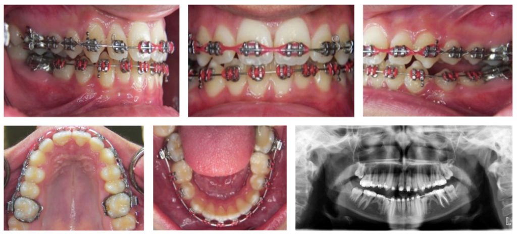
Fig 3: Clinical photographs and panoramic radiograph after a year of treatment
The total treatment duration was 24 months. Before the fixed appliances were debonded, a good functional occlusion had been established. The lower left deciduous second molar exfoliated because of root resorption. Hawley retainers were used for retention; the upper Hawley retainer included a biteplate to control vertical growth, while the lower Hawley retainer had an acrylic tooth in the edentulous space. The protocol for retention was full-time use of the retainers for 3-4 months, followed by night-time use for several years to control the vertical overlap of incisors. However, the lower Hawley retainer was to be used full time until he was ready for replacement of the missing second premolars with implants. The treatment aims were accomplished successfully
Fig 4: Post-treatment clinical photographs and panoramic radiograph

The deep bite was corrected, and normal overjet and overbite were achieved. The canine relationship was corrected to a class I relationship, the Angle class I molar relationship was maintained, and the midline of the upper and lower arches was corrected. The panoramic radiograph showed minimal bone loss in both jaws, and vertical bone loss at the distal surface of the lower left first molars. The distal root of the lower right deciduous molar was resorbed, but the tooth was still stable with no mobility. Our aim was to retain the deciduous tooth for as long as possible until it could be replaced with an implant (Figure 4). The patient exhibited
Fig 5: Clinical picture of the foot and hands showing hyperkeratosis of the palms and soles

hyperkeratosis of the palms of the hand and the soles of the feet, but these skin lesions subsided with age (Figure 5). Post-treatment evaluation after 3 years revealed some relapse of the overbite because the patient was not cooperative in wearing the retainers. An OPG revealed no bone loss compared with the previous OPG after debonding. The space of tooth #35 had decreased as a result of drifting of the lower left first molar (Figure 6).
Fig 6: 3-year post-treatment clinical photographs and panoramic radiograph

CASE 2
A female patient aged 8 years diagnosed with PLS was referred to the periodontics clinic by her primary care practitioner. She was the second child of consanguineous unaffected parents. Her elder brother also had PLS, but the other three siblings were free of apparent genetic defects. The patient’s medical history revealed that desquamation and erythema of the palms and soles were observed at the age of 4 months, with a gradual onset, followed by thickening and fissuring of both the soles. These symptoms worsened with age. The patient presented at the dermatology clinic at the National Guard Hospital, Riyadh, Saudi Arabia, when she was 2 years old with a chief complaint of palmoplantar hyperkeratosis. All laboratory tests, including hematological and liver function tests, were normal. Various therapies were undertaken to treat the skin lesions with no improvement.
Accordingly, the patient was referred to the pediatric genetic department for further investigation. The dental history revealed that during the first year after eruption of the incisors, the gingiva had become inflamed and swollen with a purulent discharge in response to the pressure of mastication. The deciduous teeth erupted with normal sequence and timing. At the age of 4 years, all the deciduous teeth were extracted under general anesthesia, aiming for complete recovery of the gingiva to its normal healthy status. Taking into consideration the clinical features, laboratory investigations, and genetic testing, a diagnosis of PLS was confirmed by the patient’s pediatrician.
The same protocol for periodontal management as for the previous patient was followed. The patient was subsequently referred to the orthodontic clinic. Intraoral examination revealed gingival inflammation, and no tooth mobility. She was in the mixed dentition stage, with a deep overbite (more than 50%) and a lack of space for the permanent dentition. A panoramic radiograph revealed space loss and mesial drifting of the first molars (Figure 7). At the
Fig 7: Pretreatment clinical photographs and panoramic radiograph

age of 9 years space maintainers were used in the form of a lower lingual arch and an upper Nance appliance to maintain the space for the permanent teeth to erupt and to prevent further movement of the posterior teeth into the edentulous area. Regular checks and follow-ups were maintained every 3-6 months until the permanent teeth erupted. At the age of 14 years, the patient presented with class I malocclusion with a skeletal class I pattern and a normal vertical relationship, with mild crowding in the upper arch and moderate crowding in the lower arch, a Class I molar and canine relationship, and a 2-mm lower midline shift to the left side. The panoramic radiograph revealed generalized horizontal bone loss, a vertical pocket distal to the lower right and upper left first molars, and mesial drifting of the lower Left first molars. She was in the permanent dentition stage, with all teeth erupted except the lower second premolars and third molars. Root dilaceration was present in many of
Fig 8: Pretreatment cast photographs and panoramic radiograph before start of orthodontic treatment

the teeth, but the dilaceration was more severe in the right upper lateral and right lower lateral teeth as compared with upper second premolars and upper left lateral incisor. The lack of space for the lower second premolars was readily detected, with -5 mm of crowding in the lower dentition
Fig 9: Clinical picture of the Face showing hyperpigmentation around the mouth, and Palm-planter hyperkeratosis of the hand and foot

and +3 mm of spacing in the upper dentition (Figure 8). The patient exhibited hyperkeratosis of the palms and soles, and dryness and around the mouth (Figure 9).
ORTHODONTIC OBJECTIVES AND MANAGEMENT
The objectives of the treatment were to relieve the crowding in the lower arch, create space for the permanent teeth, achieve class I canine and molar relationships, close spaces, and stabilize the occlusion. Preadjusted edgewise fixed orthodontic appliances (0.22″ slot Roth prescription) were bonded in the lower jaw, and then the upper jaw. A very light force was used throughout the orthodontic treatment. Initial alignment and leveling were achieved with a maxillary 0.016″ Cu-NiTi archwire, followed by 0.016, and 0.016 ×0.022 SS wire. The main problems in the lower jaw were moderate crowding and deficient space for the lower second premolars. From the periodontal viewpoint, the erupted teeth exhibited minimal bone loss, except for the lower right and left first molars, which had distal vertical pockets. Hence, the planned treatment goals were expanding the arch and creating enough space for eruption of the premolars, and stabilizing the occlusion. The spaces for the lower first premolars were achieved with interdental stripping and push coil springs (Figure 10). A class II elastic was used to correct the occlusion on the left side, and anterior cross elastics were applied to correct the lower midline. The OPG
Fig 10: Clinical photographs and panoramic radiograph after a year of treatment

revealed no bone loss and no further increase in the distal vertical pocket of the lower first molars.
The total treatment duration was 15 months. The treatment goals were accomplished, and a good functional occlusion was established. The treatment resulted in a normal overjet and overbite, and class I canine and molar relationships. A panoramic radiograph showed no or only minimal bone loss (Figure 11). During the whole period of orthodontic treatment, the patient’s periodontal status was regularly evaluated for bleeding on probing, attachment loss, and pocket depth.
Fig 11: Post-treatment Clinical photographs and panoramic radiograph

Fig 12: 3-year post-treatment panoramic radiograph

Retention was accomplished by using a fixed lingual canine-to-canine retainer in the lower arch and a Hawley retainer in the upper arch. A 3-year post-treatment OPG was taken to document the stability of the periodontal condition, and showed no further resorption of bone (Figure 12).
DISCUSSION
The etiology of the periodontal problems in PLS patients is not clearly understood. The increased prevalence of periodontal disease in PLS patients gives credence to the hypothesis that an underlying immunological deficiency is an important etiological factor causing periodontitis in PLS patients.7
This results in a reduced host response against plaque bacteria and increased susceptibility to infection.19 Aa and Gram-negative bacteria were detected in subgingival plaque, and were found to play a significant role in the etiology of PLS periodontitis.6 Patients with affected periodontal tissue are at high risk of further breakdown and loss of teeth.7
Early diagnosis and proper management of periodontal problems helps to minimize periodontal deterioration and the undesirable sequelae of the syndrome.
Periodontal therapy includes mechanical debridement by scaling and polishing, systemic antibiotics to eliminate the pathogen reservoir, extraction of hopeless mobile teeth, maintenance of good oral hygiene, and regular monitoring and recall appointments.12,19 Eradication of subgingival Aa and maintenance of good oral hygiene are key factors in in preserving permanent teeth in young PLS patients.18,20 In our reported cases, the therapeutic approaches for the primary dentition period were different. In the first case, all the primary teeth except the lower second molars exfoliated several months after eruption. A combination of systemic amoxicillin and metronidazole in addition to the maintenance of good oral hygiene was followed; this has been successful in some cases.5
Another therapeutic approach to PLS, which was followed in the second reported case, was to eradicate the pathogenic periodontal flora by extracting all the primary teeth before eruption of the permanent teeth, combined with systemic antibiotic treatment to create a safe environment for eruption of the permanent teeth. The edentulous period combined with meticulous oral hygiene determines the treatment outcome.20
After the edentulous period, the permanent teeth erupt without any guidance, and this can lead to loss of space, crowding, and collapse of the dental arch.15 The cases reported here have demonstrated the potential for successful orthodontic treatment of PLS patients under a controlled regime of periodontal treatment. Orthodontic treatment was performed to expand the arch, create space for the normal eruption of the permanent teeth, and stabilize the occlusion.
This treatment helps patients to gain a normal facial appearance rather than the collapsed appearance caused by early loss of teeth.15,20 Orthodontic treatment in PLS patients with periodontal disease poses a high risk of exacerbating periodontal breakdown and tooth loss.13 In the literature, information about orthodontic treatment in PLS patients is limited. However, there are several reported cases in which follow-up of well-planned orthodontic treatment combined with a periodontal regimen resulted in successful maintenance of a healthy dentition.15-17 In the present case reports, the patients were in the late mixed dentition period with mild to moderate crowding along with mild generalized bone loss and a deep overbite. A deep overbite is traumatic to gingival tissue, and could cause occlusal trauma to the upper anterior teeth and loss of periodontal support.21,22 Light force and moderate orthodontic tooth movements of up to 3 mm were used over the entire treatment period with good maintenance of oral hygiene. The treatment resulted in well-aligned arches with acceptable occlusion and no or only minimal bone loss. The panoramic radiographs taken at the end of treatment showed a stable periodontal condition with no bone loss. Evaluation at 3 years post-treatment revealed good stability with retention. In the first case, there was some relapse of the overbite because the patient was not cooperative in wearing the retainers, or because
vertical growth continued into the late teens. However, in the second case, the result was stable.
These case reports showed the need to discuss the possibility of orthodontic tooth movement in patients with PLS. More research and case reports are needed to document treatment in patients with PLS of varying severity levels. However, the present case reports revealed that orthodontic tooth movement is possible in patients with PLS within a combined interdisciplinary treatment protocol.
CONCLUSION
The present case reports demonstrate that PLS patients can undergo successful combined orthodontic treatment and strict periodontal therapy to achieve moderate orthodontic tooth movement. Such a combined interdisciplinary treatment regimen could be crucial in achieving functional and esthetically pleasing dentition in patients with PLS.
ACKNOWLEDGEMENTS
The author appreciatively acknowledges Prof. Nahid Ashri, Consultant Periodontist, Department of Periodontics and Community Dentistry, College of Dentistry, King Saud University, Riyadh, for her valuable and competent support and advice. We also thank Helen Jeays, BDSc AE, from Edanz Group (https://en-author-services.edanzgroup.com/ac) for editing a draft of this manuscript.
CONFLICT OF INTEREST
None to declare
REFERENCES
- French D, Scott H, Overall CM. Papillon-Lefèvre syndrome associated early onset periodontitis: a review and case study. J Can Dent Assoc. 1995;61:432-38.
- Sharma A, Kaur G, Sharma A. Papillon-Lefevre syndrome: A case report of 2 affected siblings. J Indian Soc Periodontol. 2013;17:373-77. https://doi.org/10.4103/0972-124X.115643
- Ullbro C, El-Samadi S, Boumah C, Al-Yousef N, Wakil S, Twetman S, et al. Phenotypic variation and allelic heterogeneity in young patients with Papillon-Lefèvre syndrome. Acta Derm Venereol. 2006;86:3-7. https://doi.org/10.1080/00015550510011619
- de Haar SF, Tigchelaar-Gutter W, Everts V, Beertsen W. Structure of the periodontium in cathepsin C-deficient mice. Eur J Oral Sci. 2006;114:171-73. https://doi.org/10.1111/j.1600-0722.2006.00344.x
- De Vree H, Steenackers K, De Boever JA. Periodontal treatment of rapid progressive periodontitis in 2 siblings with Papillon-Lefèvre syndrome: 15-year follow-up. J Clin Periodontol. 2000;27:354-60. https://doi.org/10.1034/j.1600-051x.2000.027005354.x.
- Eickholz P, Kugel B, Pohl S, Näher H, Staehle HJ. Combined mechanical and antibiotic periodontal therapy in a case of PapillonLefèvre syndrome. J Periodontol. 2001;72:542-49. https://doi.org/10.1902/jop.2001.72.4.542
- Hart TC, Shapira L. Papillon-Lefèvre syndrome. Periodontol 2000. 1994;6:88-100. https://doi.org/10.1111/j.1600-0757.1994.tb00029.x
- Hart PS, Zhang Y, Firatli E, Uygur C, Lotfazar M, Michalec MD, Marks JJ, Lu X, Coates BJ, Seow WK, Marshall R. Identification of cathepsin C mutations in ethnically diverse Papillon-Lefèvre syndrome
patients. J Med Genet. 2000;37:927-32. https://doi.org/10.1136/jmg.37.12.927 - Toomes C, James J, Wood AJ, Wu CL, McCormick D, Lench N, Hewitt C, Moynihan L, Roberts E, Woods CG, Markham A. Loss-offunction mutations in the cathepsin C gene result in periodontal disease and palmoplantar keratosis. Nat Genet. 1999;23:421-24. https://doi.org/10.1038/70525
- Oguzkurt P, Tanyel FC, Büyükpamukçu N, Hiçsönmez A. Increased risk of pyogenic liver abscess in children with Papillon-Lefevre syndrome. J Pediatr Surg. 1996;31:955-56. https://doi.org/10.1016/S0022-3468(96)90420-0
- Hart TC, Hart PS, Bowden DW, Michalec MD, Callison SA, Walker SJ, Zhang Y, Firatli E. Mutations of the cathepsin C gene are responsible for Papillon-Lefevre syndrome. J Med Genet. 1999;36:881-87.
- Hattab FN. Papillon-Lefèvre syndrome: from then until now. Stomatological Dis Sci. 2019;3:1 https://doi.org/10.20517/2573-0002.2018.22
- Dhanrajani PJ. Papillon-Lefevre syndrome: clinical presentation and a brief review. Oral Surg Oral Med Oral Pathol Oral Radiol Endod. 2009;108:e1-7. https://doi.org/10.1016/j.tripleo.2009.03.016
- Nickles K, Schacher B, Schuster G, Valesky E, Eickholz P. Evaluation of two siblings with Papillon-Lefèvre syndrome 5 years after treatment of periodontitis in primary and mixed dentition. J
Periodontol. 2011;82:1536-47. https://doi.org/10.1902/jop.2011.100615 - Lux CJ, Kugel B, Komposch G, Pohl S, Eickholz P. Orthodontic treatment in a patient with Papillon-Lefèvre syndrome. J Periodontol. 2005;76:642-50. https://doi.org/10.1902/jop.2005.76.4.642
- Challa P, Gandikota CS, Tarlapally S, Rayapudi N. Combined orthodontic and periodontic therapy in a patient with Papillon-Lefèvre syndrome. Journal of Dr. NTR University of Health Sciences.
2012;1:182-86. https://doi.org/10.4103/2277-8632.102450 - AlSarheed MA, Al-Sehaibany FS. Combined orthodontic and periodontic treatment in a child with Papillon Lefèvre syndrome. Saudi Med J. 2015;36:987-92. https://doi.org/10.15537/smj.2015.8.11437
- Rüdiger S, Petersilka G, Flemmig TF. Combined systemic and local antimicrobial therapy of periodontal disease in Papillon-Lefèvre syndrome. A report of 4 cases. J Clin Periodontol. 1999;26(12): 847-854.
- 23.Yousry YM, Abd EL-Latif AE, Abd El-Gawad RY. Case Report: Clinical manifestation and dental management of Papillon-Lefèvre syndrome. F1000Res. 2018;7:1420-8. https://doi.org/10.12688/f1000research.16042.1
- Toygar HU, Kircelli C, Firat E, Guzeldemir E. Combined therapy in a patient with Papillon-Lefèvre syndrome: A 13-year follow-up. J Periodontol. 2007;78:1819-24. https://doi.org/10.1902/jop.2007.070004
- Bennett JC, McLaughlin RP. Management of deep overbite with a preadjusted appliance system. J Clin Orthod1990;24:684-96.
- Tulloch JC, Proffit WR, Phillips C. Outcomes in a 2-phase randomized clinical trial of early Class II treatment. Am J Orthod Dentofacial Orthop. 2004;125:657-67. https://doi.org/10.1016/j.ajodo.2004.02.008
Consultant and Associate Professor, Department of Pediatric Dentistry and Orthodontics, College of Dentistry, King Saud University, Saudi Arabia.
Corresponding author: “Dr. Laila Fawzi Baidas†< lbaidas@ksu.edu.sa >