One Point Fixation Versus Two Point Fixation in Zygomaticomaxillary Complex Fractures


 Muhammad Husnain Akram                  BDS, MFDRCSI 

 Muhammad Mustafa Ch                         BDS, MDS

 Armaghan Israr Mirza                            BDS, FCPS, FFDRCS

 Muhammad Usman Akhtar                    BDS, MCPS, MDS, BSc

Ammar Saeed                                          BDS, FCPS

OBJECTIVE: To compare the mean satisfaction of patients undergoing one point fixation versus two point fixation for
zygomaticomaxillary complex fractures.
METHODOLOGY: This randomized controlled trial was performed at Oral and Maxillofacial surgey department Punjab
Dental Hospital. The duration of this study was 6 months (28-8-18 to 28-2-19). After meeting the inclusion criteria 74 patients
were enrolled. Informed consent and demographic information was taken. Patients were randomly divided into two groups.
One group is treated with one point fixation method and other with two point fixation. Patient were evaluated in terms of
satisfaction of score on 3rd day, 14th day and 28th day and were recorded as per operational definitions. All the collected data
was entered and analyzed on SPSS version 20.
RESULTS: In this study the mean age of patients in one point group was 42.46±15.325 years while in two points group was
49.32±13.145 years, male to female ratio of the patients was 1.2:1. The mean PSS in one point group was 5.98±0.89 while in
two points group was 3.775±0.609 (p-value=<0.001).
CONCLUSION: One point fixation is smarter method and provide significantly better satisfaction than to two point fixation
method for ZMC fractures
KEYWORDS: Zygomaticomaxillary Complex Fractures, One Point, Two Point, Satisfaction
HOW TO CITE: Akram MH, Ch MM, Mirza AI, Akhtar MU, Saeed A. One point fixation versus two point fixation in
zygomaticomaxillary complex fractures. J Pak Dent Assoc 2021;30(2):74-80.
DOI: https://doi.org/10.25301/JPDA.302.74
Received: 16 September 2020, Accepted: 25 December 2020

Download PDF

MTA Cvek Pulpotomy Followed by Fragment Re-Attachment in Traumatized Young Permanent Maxillary Left Central Incisor – A Case Report

Abul Khair Zalan                                BDS

Khadeejah Khalil Zubairy                 BDS

Hira Zaman                                         BDS, MDS

Anser Maxood                                    BDS, FRACDS, FICD, MSc

Anika Gul                                            BDS

Miraat Anser                                       BDS

Most common form of dental trauma in children are the fractures of crown mainly in anterior teeth. A case of complicated crown fracture treated with Cvek pulpotomy using MTA followed by fragment re-attachment with careful follow-up is presented here. Tooth fragment re-attachment provides feasible conservative alternative approach to restore esthetics and tooth function. Successful outcomes have been shown by the long term follow up of the treatment with the preservation of pulp vitality and continued development of root. Clinical results have also shown good esthetics and functional results of presented technique.
KEYWORDS: Complicated crown fracture, Cvek pulpotomy, MTA, fragment re-attachment
HOW TO CITE: Zalan AK, Zubairy KK, Zaman H, Maxood A, Gul A, Anser M. MTA cvek pulpotomy followed by fragment re-attachment in traumatized young permanent maxillary left central incisor – A case report. J Pak Dent Assoc 2021;30(1):70-73.
DOI: https://doi.org/10.25301/JPDA.301.70
Received: 24 July 2020, Accepted: 18 November 2020

INTRODUCTION

Most common injuries in children and young adults are the Traumatic dental injuries (TDIs). Among all the dental injuries, luxations and crown fractures occurs more frequently. Maxillary Central incisor 80% followed by maxillary lateral incisor 20% are the commonly affected teeth because of their position in the arch.1
According to Andreason’s classification, Dental fractures involving enamel, or both dentin and enamel but without puplal exposure are termed as uncomplicated crown fractures. But when the fracture is associated with pulp exposure, it is classified as a complicated crown fracture and an endodontic treatment should be considered before the definitive treatment. Approximately 18-20% of all traumatic injuries to permanent teeth are complicated crown fractures.2 Management of coronal tooth fractures depends on multiple factors which include extent and pattern of fracture, involvement of pulp tissue, violation of biological width, fracture of alveolus, any associated soft tissue injury and presence or absence of fractured tooth segment.3
In young permanent teeth, the treatment of crown fractures with pulp exposure highly depends on extent of pulp tissue involvement, the degree of development of root and most importantly the time period between the examination and traumatic incident.3 Pulp should be preserved whenever possible in such teeth with immature roots to allow root formation and tooth maturation. Pulp capping is recommended when size of pulpal exposure is less then 1 mm2and the duration of exposure at the time of treatment should not be
more then few hours.4
Cvek pulpotomy which is a technique of partial pulpotomy, is considered as treatment of choice when pulpal exposure is more than 1mm2 and time lapse is long between the incident and examination.4 Until 1983 calcium hydroxide mixture was used as a dressing material to initiate reparative dentin formation by controlling infection and stimulating the pulp healing process.4 Over the past few decades, MTA has taken the endodontic world by its storm. It improves the healing capacity of pulp tissue as it provides good ability to seal, biocompatibility, low cytotoxicity and also induces odontoblast for forming a dentinal bridge barrier. Hence MTA is considered now as a gold standard pulp dressing material.5
Esthetic treatment options for coronal tooth fractures involves ceramic (laminated veneers, full crown) or composite restorations and re-attachment of fractured tooth fragment.6
Fragment re-attachment is the finest option if the broken tooth fragment is available and in a condition that it can be used with proper occlusion, esthetics and good prognosis.6
In 1964, Chosak and Eidelman was the first who published this technique.7
Tooth fragment re-attachment provides a more conservative, esthetic and cost effective restorative approach to restore tooth function and esthetics similar to natural teeth, thus resulting a positive psychological response in the patient.8 Throughout the literature, different preparation techniques have been described to increase the retention of broken tooth fragment mechanically such as placement of a circumferential bevel, enamel groove, external chamfer, overcontour, dentinal groove and different types of adhesive materials.9 Reis have concluded that fractures restored with no further preparation in broken fragment or in effected tooth have only 37% fracture resistance, while 60% fracture resistance is increased by introducing a buccal chamfer.
Placement of internal groove and bonding with over contouring further increased the strength of intact tooth fracture upto 90% and 97% respectively.9 Presented case describes the treatment of complicated crown fracture with Cvek’s pulpotomy using MTA followed by re-attachment of broken fragment.

CASE REPORT

A female patient of 12 years old is presented to Pediatric dental department at Pakistan Institute of Medical Sciences, Islamabad with the chief complaint of broken upper front
tooth due to trauma one hour ago. She brought the broken fragment kept in saline. Medically she was fit and well with no known medical history. The intraoral examination revealed
middle third horizontal coronal fracture of upper left central incisor with the pulp exposure of more than 1mm2. Extraoral examination showed a small laceration on the ipsilateral side of lower lip (fig a,b). Diagnosis was made after doing clinical tests and radiographic examination.Three radiographs at different vertical angulations were done to rule out any root fracture. Radiographic examination revealed complicated oblique crown fracture of involved tooth involving the pulp. There was a slightly widened lamina dura of the involved tooth, without any root fracture or any periapical radiolucency. (fig c) Cold test was done initially on the sound adjacent tooth (upper right central incisor) to evaluate the normal response 

Figure (a) preoperative intraoral picture showing fractured left maxillary central incisor and lower lip laceration (b) fractured tooth showing pulp exposure (c) Periapical radiograph showing widened lamina dura(d)Rubber dam application (e) holding tooth for preparation to prevent any tooth movement(f) internal groove preparation in the fracture segment to increase retention. (g) hemorrhage control by using moist cotton pellet (h) MTA has been placed after attaining hemostasis. (i) stabilization of fracture segment on a micro brush to facilitate handling. (J) repositioning and bonding of the fracture segment via flowable composite. (k) restoration done via packable composite.(l)postoperative clinical picture after finishing and polishing.(m) immediate postoperative radiograph.(n) follow up after 1 week. (0) last picture showing postoperative radiograph after 1 year.

by using ethyl chloride.Again it was done on upper left central incisor (traumatized tooth) which showed no response, because of the pulp shock. The tooth was not mobile but was slightly tender to percussion because of the associated subluxation and PDL injury. Lateral soft tissue radiograph of lower lip was done in order to rule out any foreign body. No foreign body was present upon radiographic evaluation. After clinical and radiographic evaluation, the definitive diagnosis cannot be done as it was traumatized 1hour ago.
The decision was made to perform cvek (partial) pulpotomy with MTA (ProRoot, Dentsply) as pulp exposure was more than 1 mm2, followed by fragment reattachment of traumatized tooth.. Broken tooth fragment was assessed intraorally for proper occlusion.
After taking an informed consent, topical anesthesia (benzocaine 20%, keystone USA) was applied after drying the oral mucosa. Local maxillary infiltration was done with 2% lidocaine (1:100,000). To prevent salivary contamination, operating field was isolated using rubber dam. (fig d) Tooth was washed with copious amount of saline. By holding the tooth, 2 mm coronal pulp tissue, below the level of pulp exposure was gently removed using sterile round bur mounted on high speed handpiece with continuous saline irrigation (fig e). Bleeding was controlled by placing sterile moistened cotton pellet. (fig f) after attaining hemostasis within 3 minutes, MTA (ProRoot, Dentsply) powder was dispensed
on a glass slab and mixed with distilled water according to the manufacturer’s recommendation and placed over the exposed pulp without any pressure . Afterward, a Resin modified Glass Ionomer Cement (FUJI IX, GC Corporation, Tokyo, Japan) was applied as a base material to seal the cavity.
Prior to re-attachment procedure sharp margins of the tooth and the broken fragment were smoothened. Using small sterile #2 round bur (R40004G, Coltene) internal groove was made on a broken fragment to increase retention (fig f). Broken fragment was then secured with the tip of bonding brush in order to facilitate handling. It is then etched by using 37% phosphoric acid (META BIOMED CO.Lt) for 30 sec followed by washing for 20 sec and drying with a moist cotton pellet. Dentine bonding agent (Meta P & Bond, META BIOMED) then applied over the etched surface and light curing was not done at this point. Likewise, fractured residual tooth surface also treated with 37% phosphoric acid etchant (META BIOMED CO.Lt) then washed and same dentine bonding agent (Meta P & Bond, META BIOMED) was applied to it. Flowable composite resin (Filtek Z350 XT, USA) was then applied to both broken fragment and fractured tooth surface. Fragment was repositioned properly on the tooth. Excessive resin was wiped off after establishing the appropriate position and light cured for 40 sec on both labial and lingual surfaces. (fig g). After assuring proper stability of the fragment, ‘double chamfer’ margin of 1mm was given using a round end tapered bur(dentsply) above and below the fracture line of the tooth. Permanent composite restoration (3M ESPE, Z250,USA) was done to restore the tooth. (fig i) It was then finished and polished. Gross contouring and finishing was initially done with tapered round ended finishing bur(DENTSPLY/Caulk). Intermediate contouring and finishing was done with soflex discs ( 3M, ESPE) then final polishing was done with diamond polishing paste (Kerr). Rubber dam was removed and occlusion was checked carefully by using an articulating paper. Post-operative instructions to abstain from applying heavy occlusal forces on this tooth was given and patient was motivated to practice good oral hygiene. Before dismissing the patient, PA radiograph of the treated tooth was done for comparison in the follow-up appointments (fig j). Careful clinical and radiographic examinations were
done after 1week, 3 weeks and 6 weeks in order to check the vitality of the tooth and root growth. Periapical radiographs showed no pathological changes at 1week, 3weeks and
6weeks.Cold test was done at each follow-up appointment which showed negative response at 1week postoperatively and showed positive response at 3 week and 6 week of
follow-up examination. (fig n) Percussion test was also done at each followup visit that showed negative response.

DISCUSSION

Complicated crown fracture are the fractures involving both dentine and enamel with pulp exposure. 18-20% of all the traumatic injuries involves these type of fractures.2 Such
fractures should be managed as early as possible to prevent pulp necrosis. To keep the pulp vital, in fractures involving pulp exposure of upto 4mm, Partial vital pulpotomy is the
treatment of choice. 96% success rate is reported by Cvek in such cases with long follow up period.10 Calcium hydroxide was previously used in vital pulpotomy. MTA is now the material of choice with reparation mechanism is similar to calcium hydroxide as it provides better long term seal and produce more dentinal bridging in relatively shorter period of time with less pulp tissue inflammation.10 Secondly, in the current case the method used to reattach the fracture segment involves preparation of the internal groove in the fractured fragment and double chamfer formation on the crown portion 1 mm above and below fracture line to improve strength and retention. One year clinical and radiographic follow up was done.
Clinical results at the end of follow up showed adequate aesthetic and functional results of fragment re-attachment technique with no pain, sensitivity, pathological pulpal changes and change of tooth color. Radiographic examination also showed no pathological periapical / peri-radicular changes, root resorption or calcification of canals.

CONFLICT OF INTEREST

None to declare

REFERENCES

  1. J. O. Andreasen, “Etiology and pathogenesis of traumatic dental injuries: a clinical study of 1,298 cases,” Scand J Dent Res. 1970; 78: 329-42. https://doi.org/10.1111/j.1600-0722.1970.tb02080.x
  2. De Blanco LP. Treatment of crown fractures with pulp exposure. Oral Surg Oral Med Oral Pathol Oral Radiol Endod. 1996;82:564-8. https://doi.org/10.1016/S1079-2104(96)80204-6
  3. Olsburgh S, Jacoby T, & Krejci I. Crown fractures in the permanent dentition: Pulpal and restorative considerations Dent Traumatol. 2002;18:103-15. https://doi.org/10.1034/j.1600-9657.2002.00004.x
  4. Cvek M. Endodontic treatment of traumatized teeth. In: Andreasen JO, editor. Traumatic injuries to the teeth. 2nd ed. Copenhagen: Blackwell Munksgaard; 2003;321-83.
  5. Ford TR, Torabinejad M, Abedi HR, Bakland LK, Kariyawasam SP. Using mineral trioxide aggregate as a pulp capping material. J Am Dent Assoc. 1996;127:1491-94. https://doi.org/10.14219/jada.archive.1996.0058
  6. Alvares I, Sensi LG, Araujo EM Jr, & Araujo E. Silicone index: An alternative approach for tooth fragment reattachment. J Esthetic Restorative Dentist. 2007;19:240-46.
    https://doi.org/10.1111/j.1708-8240.2007.00110.x
  7. CMC Taguchi, JK Bernardon, G Zimmermann, and LN Baratieri. Tooth Fragment Reattachment: A Case Report. Operative Dentistry: 2015;40;227-34. https://doi.org/10.2341/14-034-T
  8. Badami V, & Reddy SK. Treatment of complicated crown-root fracture in a single visit by means of rebonding. J Am Dent Assoc. 2011;142:646-50.
    https://doi.org/10.14219/jada.archive.2011.0246
  9. Reis A, Francci C, Loguercio AD, et al. Re-attachment of anterior fractured teeth: fracture strength using different techniques. Oper Dent 2001;26:287-94.
  10. M. Cvek, “A clinical report on partial pulpotomy and capping with calcium hydroxide in permanent incisors with complicated crown fracture,” J Endod. 1978;232-37. https://doi.org/10.1016/S0099-2399(78)80153-8

  1. MDS (Resident), Post graduate, Department of Pediatric Dentistry, Pakistan Institute of Medical Sciences.
  2. House officer, Department of Pediatric Dentistry, Pakistan Institute of Medical Sciences.
  3. Registrar, Department of Operative Dentistry, University College of Dentistry.
  4. Dean of Dentistry, Department of Operative & Pediatric Dentistry, Pakistan Institute of Medical Sciences.
  5. FCPS (Resident), Post graduate, Department of Orthodontics, Sardar Begum Dental College.
  6. MDS (Resident), Post graduate, Department of Pediatric Dentistry, Pakistan Institute of Medical Sciences.
    Corresponding author: “Dr. Abul Khair Zalan” < zalanjan@yahoo.com >

MTA Cvek Pulpotomy Followed by Fragment Re-Attachment in Traumatized Young Permanent Maxillary Left Central Incisor – A Case Report

Abul Khair Zalan                                BDS

Khadeejah Khalil Zubairy                 BDS

Hira Zaman                                         BDS, MDS

Anser Maxood                                    BDS, FRACDS, FICD, MSc

Anika Gul                                            BDS

Miraat Anser                                       BDS

Most common form of dental trauma in children are the fractures of crown mainly in anterior teeth. A case of complicated crown fracture treated with Cvek pulpotomy using MTA followed by fragment re-attachment with careful follow-up is presented here. Tooth fragment re-attachment provides feasible conservative alternative approach to restore esthetics and tooth function. Successful outcomes have been shown by the long term follow up of the treatment with the preservation of pulp vitality and continued development of root. Clinical results have also shown good esthetics and functional results of presented technique.
KEYWORDS: Complicated crown fracture, Cvek pulpotomy, MTA, fragment re-attachment
HOW TO CITE: Zalan AK, Zubairy KK, Zaman H, Maxood A, Gul A, Anser M. MTA cvek pulpotomy followed by fragment re-attachment in traumatized young permanent maxillary left central incisor – A case report. J Pak Dent Assoc 2021;30(1):70-73.
DOI: https://doi.org/10.25301/JPDA.301.70
Received: 24 July 2020, Accepted: 18 November 2020
Download PDF

A Permanent solution to Restore Occlusal Vertical Dimension and Partial Edentulism – Overlay Removable Partial Denture

Surhan Aziz                                         BDS

Muhammad Rizwan Nazeer              BDS, FCPS

Kamil Zafar                                         BDS

Robia Ghafoor                                   BDS, FCPS

Tooth wear is a common problem seen in population and poses restorative challenge to a dentist as well. Extensive dental procedures are usually required for the treatment of tooth wear associated with loss of occlusal vertical dimension. An overlay removable partial denture provides conservative treatment option for patients with moderate to severe worn down dentition and partial edentulism. It is designed in such a way that some part of denture component also covers the occlusal surface of remaining teeth and replace the remaining teeth to achieve a functionally stable occlusion. This case report is about a patient who was given cast metal overlay upper and lower denture for a correction of lost vertical dimension of occlusion, replacement of few missing teeth and for the prevention from further tooth wear.
KEYWORDS: Tooth wear, vertical dimension, overlay denture
HOW TO CITE: Aziz S, Nazeer MR, Zafar K, Ghafoor R. A Permanent solution to restore occlusal vertical dimension and partial edentulism – Overlay removable partial denture. J Pak Dent Assoc 2021;30(1):66-69.
DOI: https://doi.org/10.25301/JPDA.301.66
Received: 10 June 2020, Accepted: 24 August 2020

Tooth wear is defined as the progressive loss of tooth structure from attrition, abrasion, erosion or abfraction.1 It has a multifactorial cause and can be localized to anterior or posterior teeth or often generalized throughout the dentition.2 When the frequency of tooth wear is slow, the resultant occluso-vertical dimension is often maintained through dento-alveolar compensation.3 However, the cases of accelerated tooth wear often results in loss of occluso-vertical dimension and hence an increase in freeway space is usually found.3-4 Loss of occlusal vertical dimension (OVD) significantly affects patients quality of life by altering aesthetics and function, hence requires a systematic approach for appropriate management.5,6
The management of accelerated tooth wear is often complicated by patient complains (e.g pain and sensitivity), which requires immediate treatment to relief symptoms. Following that, a preventive approach is employed for preventing further tooth loss.6 Definitive treatment plan is then aimed at regaining the lost occlusal vertical dimension, with fixed prosthodontics being the most preferred option.7 Extensive dental procedures such as multiple endodontics, post core, crown lengthening surgeries are usually required before fixed prosthodontics, which not only results in irreversible loss of pulp vitality but also a great deal of financial expenditure and time to achieve desire results.4,5,7,8 An overlay removable partial denture is a conservative treatment option for patients with moderate to severe worn down dentition and partial edentulism.9
It is designed in such a way that some part of denture component also covers the occlusal surface of remaining teeth and replace the missing teeth to achieve a functionally stable occlusion.9,10 It can also be used as transitional prostheses prior to fixed full mouth rehabilitations or in patients with severe skeletal deficiency such as ectodermal dysplasia, Kenny-Caffey Syndrome 11 and malocclusion, but can be considered as or as permanent prosthesis in severe tooth wear cases and in patient with financial and/or time constraints.4,12
The present case report is about a patient who was given cast metal overlay bimaxillary dentures for a correction of lost vertical dimension of occlusion, for replacement of few missing teeth and for the prevention from further tooth wear.

CASE PRESENTATION

A 53 year old male presented to the dental clinics of Aga Khan University Hospital, Karachi Pakistan with the complaint of extreme sensitivity in lower left jaw for several months. Pain aggravated on taking hot and cold beverages but since the last one week it also increases on taking room temperature water and lingers for more than few minutes after removal of the stimulus. The patient’s medical history was insignificant, except hypertension for which he was on medications. His last visit to a dentist was 3 to 4 years ago for extraction of teeth on right side.
On extra oral examination, no swelling or lesions were present and findings for lymphadenopathy were negative. Muscles of mastication were tender to palpation and TMJ
examination revealed no clicking or crepitus. Range of mandibular movements was within normal limits and freeway space was found to be 6 mm with reduced occlusal vertical dimension (OVD). On intraoral examination, soft tissues were normal. Teeth # 17,16,47,46 were absent and generalized moderate to severe tooth wear was found. Increased overjet
and overbite were also observed. Radiographs and pulp vitality test was performed for left lower teeth which revealed irreversible pulpitis in tooth # 37 and reversible pulpitis in
# 36. Extra- and intra-oral photographs were initially taken for case assessment. (Figure 1 & 2) Questions regarding stress, lifestyle, diet, medications, parafunction, brushing habits frequency of acidic drinks were also inquired. Para

Figure 1: Pre-operative intra-oral pictures

Figure 2: Pre-operative OPG Xray

functional habit (clenching) was positive in our patient. Study casts were also obtained. On the basis of thorough clinical and radiographical examination, problem list was consisted of:

  1. Moderate to severe tooth wear with reduced occlusal vertical dimension of occlusion (freeway space =6 mm) without dentoalveolar compensation.
  2. Irreversible puplitis # 37
  3. Reversible pulpitis # 36
  4. Missing teeth # 16, 17, 46, 47.

The treatment plan comprised of:
Immediate management: Root canal treatment # 37 and composite resin restoration # 36 for the relief of pain and sensitivity. Patient was also provided stabilization splint to
prevent further wear and establish new OVD. Control phase: Topical fluoride and oral hygiene instructions were given and after 3 months patient was evaluated for definitive management. Definitive phase: Placement of indirect full coverage restorations on all posterior teeth at an increased occlusal height, implant retained crown for missing teeth and metal palatal veneers on maxillary anterior teeth vs placement of an overlay partial denture. Monitoring: Regular follow up were advised to evaluate the timely status of tooth wear.
Initially, emergency treatment of sensitivity was carried out which included endodontic treatment in tooth # 37 for relief of patients symptoms and canals were obturated upto
length. Composite restoration (3M Filtek P-60, 3M ESPE) was then placed in # 36. Impressions were obtained with irreversible hydrocolloid and patient was provided stabilization split which served two purposes. One was to prevent further progression of tooth wear and other was to establish new OVD for the accommodation of definitive prosthesis. All treatment options regarding prosthetic rehabilitation and pros and cons of each was thoroughly discussed with the patient. Patient was asked to think over all the options before selecting the final treatment option. A daily application of sodium fluoride 1.1% gel was advised for management of generalized sensitivity.
At the later appointment, splint was adjusted and delivered to the patient. All treatment options were re-discussed. Since the patient had financial constraints and wanted the treatment in a shorter time period, hence opted for overlay removable partial denture. After 3 months mouth preparation was done and impression was obtained with aqusail soft putty regular set (Dentsply Middle east and Africa) for the fabrication of overlay removable partial denture. At the subsequent appointment, framework trial (with acrylic teeth replacing was performed) was carried out. The framework was passive fit and comfortable to the patient. Wax bite was taken with Rigisel 2X VPS bite registration material (Dentsply Middle east and Africa) directly from teeth and mounted on semi adjustable articulator (Hanau Articulator, Teledyne Hanau Buffalo, NY, USA) in the subsequent appointment. Patient’s feedback was obtained regarding increase in occlusal vertical dimension. After patients approval the denture was finally processed in acrylic. The final denture was delivered and occlusion was checked (Figure 3). Reorganized occlusion was successfully achieved as planned. Post-operative instructions regarding

Figure 3: Post-operative pictures

denture wearing, hygiene and food intake was given to the patient. Patient was asked to follow up after 24 hours. Mild discomfort was reported by the patient so muscle relaxants
and analgesics were given for next three days. On a follow up visit after a week patient was doing absolutely fine with no discomfort. Patient was asked to wear prosthesis initially
for all the time even during the sleep. After the patient got accustomed to the prosthesis, he was advised to wear the for day time only. Next follow up was planned after 6 months.

DISCUSSION

The multifactorial etiology of tooth wear demands thorough history, clinical and radiographical examination together with special tests (TMJ screening, vertical dimension of occlusion, vitality tests) and occlusal cast examination before reaching the final diagnosis.10,11 Such patients usually presents with pain and sensitivity at advance wear stage,
making the pathologic condition more complex.7
Those presenting with lost occluso-vertical dimension and partial edentulism often poses a great restorative challenge to a dentist.7,12 The management plan should be aimed at relieving acute patients complains, preventing further tooth wear, definitive prosthetic management which should be in the best interests of the patients and frequent monitoring
throughout the treatment phase and afterwards.5,13 The treatment should be conservative, cost effective, comfortable and aesthetically pleasing to the patient.14,15 The initial management could vary depending on the clinical presentation such as from simple smoothening of a sharp edges of teeth, managing sensitivity by applying desensitizing agent or glass ionomer cement over exposed dentine.14 In cases with pulp exposure root canal treatment or dental extraction may be required in cases with severe tooth wear.14 We also followed a systematic approach in our patient, carried out endodontic treatment for relief of acute pain and also covered the exposed dentinal tubules with composite resin restoration for management of acute sensitivity in lower first molar. We also advised fluoride gel for management of generalized sensitivity and once the acute complained were settled, definitive prosthetic phase was initiated.
Depending upon the extent of tooth wear and associated loss of vertical dimension of occlusion, a patient can be categorized and are managed accordingly.7 Our patient had loss of occlusal vertical dimension with moderate tooth wear and wanted the most conservative, reversible and cost effective option. Therefore we planned an overlay denture because a single overlay denture can address multiple situations at a time such as it covers occlusal surfaces of teeth hence increase OVD, also protects teeth from further wear and sensitivity and can also replace missing teeth and porcelain veneer can also be applied on the metal framework covering anterior teeth.8-12,15-17 The other benefit of overlay denture is preservation of alveolar bone, as opposed to complete denture, therefore provides proprioception in patients with para-functional habits.15,16 Since the tooth wear was extensive in posterior teeth therefore the overlay denture covered the occlusal surface of all posterior teeth, replace the missing teeth and covered only the palatal surface of anterior teeth.
Despite multiple advantages of an overlay dentures, there are few disadvantages like compromised esthetics encountered by patients on removal of the prosthesis, increase food impaction and difficulty in maintaining oral hygiene which may lead to increased dental issues like caries and periodontitis.17 Since the overlay denture was not covering the anterior teeth in our patient hence the final outcome was aesthetically pleasing and comfortable to the patient. It is essential for the management of tooth wear that emphasis should be given to regular recall and maintenance visits to ensure the long term success of overlay partial dentures.4 We also did periodic follow-ups after delivery of final prosthesis on day one, after a week and a month. Next follow-up is planned after six months.

CONCLUSION

The purpose of the present case is to highlight the use of overlay removable partial dentures (ORPD) for the correction of lost occlusal vertical dimension (VDO) in a patient with moderately worn dentition, and partial edentulism. It is a conservative treatment option in cases with low esthetic concerns, if patients wish for simple procedures, limited finances or certain debilitating medical limitations.

CONFLICT OF INTEREST

None declared

REFERENCES

  1. Donachie MA, Walls AW. The tooth wear index: a flawed epidemiological tool in an ageing population group. Community Dent Oral Epidemiol. 1996; 24:152-8. https://doi.org/10.1111/j.1600-0528.1996.tb00833.x
  2. Faigenblum M. Removable prostheses. Brit Dent J. 1999;186: 273-76 https://doi.org/10.1038/sj.bdj.4800086
  3. Zengingul A, Eskimez S, Deger Y, Kama J. Tooth wears and dentoalveolar compensation of vertical height. Biotechnol Equip. 2007;21:362-65. https://doi.org/10.1080/13102818.2007.10817474
  4. Patel MB, Bencharit S. A treatment protocol for restoring occlusal vertical dimension using an overlay removable partial denture as an alternative to extensive fixed restorations: a clinical report. Open Dent J. 2009;3:213-18 https://doi.org/10.2174/1874210600903010213
  5. Smith BG, Bartlett DW, Robb ND. The prevalence, etiology and management of tooth wear in the United Kingdom. J Prosthet Dent. 1997;78:367-72. https://doi.org/10.1016/S0022-3913(97)70043-X
  6. Bartlett DW. The role of erosion in tooth wear: aetiology, prevention and management. Int Dent J. 2005;55:277-84. https://doi.org/10.1111/j.1875-595X.2005.tb00065.x
  7. Turner KA, Missirlian DM. Restoration of the extremely worn dentition. J Prosthet Dent. 1984;52:467-74. https://doi.org/10.1016/0022-3913(84)90326-3
  8. Windchy AM, Morris JC. An alternative treatment with the overlay removable partial denture: a clinical report. J Prosthet Dent. 1998; 79:249-53. https://doi.org/10.1016/S0022-3913(98)70232-X
  9. Beyth N, Tamari I, Buller Sharon A. Overlay removable denture for treatment of worn teeth. Spec Care Dentist. 2014; 34:295-97. https://doi.org/10.1111/scd.12063
  10. Bataglion C, Hotta TH, Matsumoto W, Ruellas CV. Reestablishment of occlusion through overlay removable partial dentures: a case report. Braz Dent J. 2012; 23:172-74.
    https://doi.org/10.1590/S0103-64402012000200014
  11. Demir T, Kecik D, Cehreli ZC. Kenny-Caffey Syndrome: Oral Findings and 4-year Follow-up of Overlay Denture Therapy. J Dent Child. 2007;74:236-40.
  12. Nosouhian S, Davoudi A, Derhami M. Posterior open occlusion management by registration of overlay removable partial denture: A clinical report. J Indian Prosthodont Soc. 2015;15:386-89 https://doi.org/10.4103/0972-4052.171822
  13. Bloom DR, Padayachy JN. Increasing occlusal vertical dimension- -why, when and how. Br Dent J. 2006;200:251-56 https://doi.org/10.1038/sj.bdj.4813305
  14. Mehta SB, Banerji S, Millar BJ, Suarez-Feito JM. Current concepts on the management of tooth wear: part 1. Assessment, treatment planning and strategies for the prevention and the passive management of tooth wear. Br Dent J. 2012;212:17-27. https://doi.org/10.1038/sj.bdj.2011.1099
  15. Ganddini MR, Al-Mardini M, Graser GN, Almog D. Maxillary and mandibular overlay removable partial dentures for the restoration of worn teeth. J Prosthet Dent 2004;91:210-14. https://doi.org/10.1016/j.prosdent.2003.12.021
  16. Beumer III J, Hamada MO, Lewis S. A prosthodontic overview. Int J Prosthodont. 1993;6:126-30 https://doi.org/10.1097/00008505-199312000-00009
  17. Del Castillo R, LaMar F, Ercoli C. Maxillary and mandibular overlay removable partial dentures for the treatment of posterior openocclusal relationship: a clinical report. J Prosthet Dent. 2002;87:587-92. https://doi.org/10.1067/mpr.2002.125578

  1. Post Graduate trainee Department of Operative Dentistry at the Aga Khan University Hospital, Karachi, Pakistan
  2. Senior Registrar Department of Operative Dentistry at the Baharia University Medical and Dental College, Karachi, Pakistan.
  3. Instructor, Department of Operative Dentistry at the Aga Khan University Hospital, Karachi, Pakistan
  4. Assistant Professor, Department of Operative Dentistry at the Aga Khan University Hospital, Karachi, Pakistan.
    Corresponding author: “Dr. Robia Ghafoor” < robia.ghafoor@aku.edu >

A Permanent solution to Restore Occlusal Vertical Dimension and Partial Edentulism – Overlay Removable Partial Denture

Surhan Aziz                                         BDS

Muhammad Rizwan Nazeer              BDS, FCPS

Kamil Zafar                                         BDS

Robia Ghafoor                                   BDS, FCPS

Tooth wear is a common problem seen in population and poses restorative challenge to a dentist as well. Extensive dental procedures are usually required for the treatment of tooth wear associated with loss of occlusal vertical dimension. An overlay removable partial denture provides conservative treatment option for patients with moderate to severe worn down dentition and partial edentulism. It is designed in such a way that some part of denture component also covers the occlusal surface of remaining teeth and replace the remaining teeth to achieve a functionally stable occlusion. This case report is about a patient who was given cast metal overlay upper and lower denture for a correction of lost vertical dimension of occlusion, replacement of few missing teeth and for the prevention from further tooth wear.
KEYWORDS: Tooth wear, vertical dimension, overlay denture
HOW TO CITE: Aziz S, Nazeer MR, Zafar K, Ghafoor R. A Permanent solution to restore occlusal vertical dimension and partial edentulism – Overlay removable partial denture. J Pak Dent Assoc 2021;30(1):66-69.
DOI: https://doi.org/10.25301/JPDA.301.66
Received: 10 June 2020, Accepted: 24 August 2020
Download PDF

Leukemia and Periodontal Health

Bann AlHazmi                    BDS, MSc

Leukemia is a malignant neoplasm that arises from hematopoietic cells. leukemia has high prevalence among Saudi and Pakistani populations (6.2% and 4.1% respectively). Dentists should perceive the serious complications of leukemia and its therapies and to manage leukemic patients in dental practice safely and effectively. Oral manifestations of hematological malignancies may represent the initial sign of the underlying hematopoietic disease. 65% of leukemia lesions have some form of oral manifestations. Accordingly, the purpose of this review is to summaries all the general manifestations of Leukemia as well as the oral manifestations to facilitate early diagnosis and referral. Leukemia oral manifestations could be pale mucosa or spontaneous bleeding gingiva or bruising and petechiae in the hard and soft palate. Gingival hyperplasia, ulcerations, and opportunistic infections with Candida albicans and Herpes Viruses can occur in oral mucosa.
Dental care for leukemic patients should focus on trauma prevention and meticulous oral hygiene. Antibiotics should be used to prevent and to treat infections (bacterial, virus and fungal infections). Periodontal surgeries like gingivoplasty procedures are considered elective treatments before the diagnosis and or treatment of leukemia and it should not be performed until the patient completes and maintains their antineoplastic treatment. Dentists plays an essential role in the early diagnosis of the leukemia; therefore, they should lead a proper investigation and referral to reach proper diagnosis.
KEYWORDS: Gingival Bleeding, Leukemia, Oral Manifestations, Periodontal Surgeries.
HOW TO CITE: AlHazmi BA. Leukemia and periodontal health. J Pak Dent Assoc 2021;30(1):61-65.
DOI: https://doi.org/10.25301/JPDA.301.61
Received: 12 August 2020, Accepted: 27 November 2020

INTRODUCTION

There are many types of malignant tumors and Leukemia is one of them. Leukemia originates from abnormal hematopoietic cells, with abnormal function, and it has different types. In 2018, the number of Leukemic patients around the world reached 437, 033 cases and it is expected to rise to 23.6 million case per year by 2030.1
In Saudi Arabia, leukemia is ranked the 5th among all cancers (7.6% in males and 4.4% in females).2 The prevalence of children (age less than 14 years) with Leukemia
is 38.8% in 2017. Leukemia has the highest prevalence in Saudi children among other cancers.2 Leukemia also ranked the 5th most frequent cancer in Pakistan by new cases and
deaths, and it constitutes about 4.1% of total cancer cases (in both sexes and all ages) in 2018.3
The five-year prevalence for leukemic cases in Pakistan is 15980, and the male patients are more prevalent than female (59.7% and 40.3% respectively).3 This high leukemic prevalence should draw the attention of health care providers. It is recommended for dentists to perceive the serious complications of leukemia and its therapies and to manage leukemic patients in dental practice safely and effectively. The US National Cancer Institute4, necessitates to manage leukemic patients with multidisciplinary approach. The multidisciplinary therapy team should have professionals from oncology, radiation, nutritional, dental, hematology, social and other departments.
The dental professionals (generals and specialists) are important to diagnose and treat and prevent oral complications in leukemic patients. Oral manifestations of hematological malignancies could be the first sign of the underlying hematopoietic abnormalities. Stafford et al. reported that early diagnosis of large numbers of acute non-lymphocytic leukemias cases was a contribution of dentists.5 65% of leukemias have some form of oral pathology manifestations.5
The common general manifestations clinically are fatigue, weight loss, night sweats, pyrexia, anorexia, pallor, pruritus, splenomegaly, and hepatomegaly.6 Accordingly, the purpose of this review is to summaries all the general manifestations of Leukemia as well as the oral manifestations to facilitate early diagnosis and referral.

ORAL MANIFESTATIONS

Orofacial manifestations, especially in acute leukemia cases, have clinical appearance basically due to leukemic cells infiltration into the oral soft and hard tissues and bone marrows. The side effects of leukemia treatments are considered secondary and tertiary effects. For example, recurrent aphthous-like ulcers and periodontal disease are
mostly associated with qualitative defects in granulocytes. This defect is one of the chemotherapy side effects.
Oral mucosa: the oral mucosa could look pale or erythematous. The pale mucosa is due to anemia. Bleeding of oral mucosa and blue petechia seen in the palate, tongue or lips are common. The mucosal bleeding is a result of thrombocytopenia.7 Ulcerations and infections with Candida albicans and Herpes Viruses can occur in oral mucosa because of immunosuppression condition.8
Gingiva: gingival hyperplasia especially in interdental papilla or gingival margins is common in acute cases. The hyperplasia is a result of inflammation or leukemic cell infiltration and it could be localized or generalized.9 On the other hand, chronic leukemic cases have lesser infiltration and the gingiva would be paler.9 Severe thrombocytopenia (the platelet count is lower than 20,000 cells/mm3) can cause gingival bleeding.10 However, if the thrombocytopenia platelet count is above 20,000 cells/mm3, gingiva and palate manifest bruising and petechiae.10
Teeth: changes in teeth are not a primary cause changes but a treatment side effects. Inability to maintain good oral hygiene, tendency to eat soft diet and changes in oral microflora habitat are the main reason of cervical teeth caries. Bone leukemic cell infiltration mostly in acute cases, is responsible for teeth displacement and destruction of alveolar
bone and periodontal ligaments.5,11 Another possible manifestation is tooth pain, and it is due to pulp infiltration.
Bone: bony masses in mandible and maxilla can occur in leukemic patients, however, their incidence is rare.12 There are four reported cases with bony masses in oral cavity, three
are B-lymphoblastic lymphoma and one T-lymphoblastic leukemia/lymphoma.13 These bony masses may look as one hard swelling or multiple lobulated friable mass. On X-rays, these bony masses appear as lytic or sclerotic bone alterations and resemble bone tumors.13 Leukemic patients may experience pain or paresthesia in the jaws area. Radiographically, there are loss of lamina dura, changes in periodontal space, resorption of the alveolar bone, and destruction of the bone structure.14 All these symptoms are
related to leukemic cell infiltrations. There are secondary and Tertiary complications related to leukemias. The side effects of antineoplastic treatments like radiation therapy or chemotherapy are considered secondary complications. Examples of secondary complications are the complications related to thrombocytopenia, granulocytopenia, and anemia.15 The tertiary complications are due to an interaction between therapy itself, the therapy’s side effects, and a systemic condition emerges from that therapy. Taste alteration, skin
desquamation, candidiasis, xerostomia, dysphasia, opportunistic infections, permanent taste loss, salivary flow reduction, osteoradionecrosis, chondronecrosis and trismus are examples of tertiary complications.15

MANAGEMENT OF LEUKEMIC ORAL MANIFESTATIONS

Treatment of oral manifestations in patients with leukemia is complicated and depends on case stability and prognosis of the disease. Therefore, dental management for leukemic
patients should focus on trauma prevention like traumas from ill-fitting dentures or faulty fixed prosthesis, bleeding elimination and prevention and treatment of any oral infection
related to leukopenia. Meticulous oral hygiene practices (teeth brushing, fluoride application, and low-sugar food) should also be stressed before, through and after treatment.16
For example, the manifestation of gingival enlargement is managed by meticulous oral hygiene, use of soft toothbrush and antiseptic mouth wash like chlorhexidine 0.12% twice
daily. Oral ulceration can be treated with topical corticosteroid gel (fluocinonide 0.1%, Apply thin layer each day or every 12 hours) and biopsy if necessary. Antibiotic is recommended for ulcers to prevent bacterial infections. Secondary infections are common; therefore, antibiotics should be administered in therapeutic rather than prophylactic doses.16,17 Antifibrinolytic mouth rinse is recommended to manage gingival bleeding, as well as good plaque control.18 All infections foci must be eradicated completely, by antibiotics or antivirals or antifungals as needed. These medications are to be used as prevention or treatment.18

General Considerations Regarding Dental Management in Leukemic patients
Dental management for leukemic patients is provided through three different stages:

1) pre-antineoplastic treatment assessment and preparation of patients,

2) oral health care during treatment,

3) post-treatment care.9,19

Pre-Antineoplastic Treatment Assessment and Patient Preparation:
Dental care in this stage is directed to treat emergencies and urgent needs only. Elective treatment should be suspended until the case gets stable clinically and laboratory. It is quite
important to educate leukemic patients or their relatives the practice of meticulous oral hygiene and how it is always essential to reduce oral problems and discomfort.4,18 Also, patients must be informed about possible side effects that may arise from antineoplastic therapy in oral cavity such as mucositis and oral pain. Dentists should focus on prevention of oral infections and injuries like sharp denture edges and orthodontics injuries. Treatment of oral mucosa lesions, carious teeth, endodontic and periodontal diseases, replacing teeth with good fitting dentures and temporomandibular joint dysfunction are examples of treatment that can be done at this stage.4
Oral Health Care during Antineoplastic Treatment:
Patients in this stage are classified as high-risk, because of infections possibility and its serious consequences in patients’ immunosuppressed condition. Any oral infection can be fatal or at least worsen the general health status of patients.20 The main goals of any dental management at this stage are to keep optimal oral health, to treat the therapy side effects, and to eradicate oral infections.4 In this stage there are common chemotherapy side effects. Main oral side effects could be high caries rate, gingival bleeding, gingival abscess, salivary gland dysfunction, herpetic gingivo stomatitis, xerostomia.4 There is no restriction to do dental examination, radiographs, prophylaxis and supragingival scaling during antineoplastic treatment phase.17
Post-Antineoplastic Treatment Oral Health Care:
After the completion of leukemia therapy, patients can be treated normally. Patients completed their antineoplastic therapy, and usually there are no oral manifestations neither
from illness nor from treatment. However, children who received chemotherapy during tooth formation, could present with enamel demineralization and abnormal dental roots.15
Elective dental treatment regimens are allowed.17 However, antibiotics prophylaxis is mandatory for at least six months post-treatment. All surgical dental procedures should be covered with antibiotics prophylaxis.21 Orthodontic therapy is allowed only after two years of post-therapy and cancer free condition.17 The dental treatment must be planned according to the disease and therapy status. Patients in good status with acceptable immune condition can receive more invasive dental procedure. However, the invasive procedures must be done after consultation with hematologist, and platelet count and neutrophil count laboratory tests must be evaluated.9 Periodontal Procedures at Different Stages of Leukemic therapy
The periodontal therapy should aim to treat and avoid any infections, or bleeding. It is of quite importance to consider some hematological indices like neutrophils and platelets counts before any periodontal procedure. Noninvasive procedures do not require special precautions and can be done at any stage of the therapy or disease. For example, clinical examination, radiographic imaging, oral hygiene practices, supragingival scaling and prophylaxis, are noninvasive procedures.17 According to Haytac et al22 performing periodontal
probing, root planing or extractions requires a neutrophil number of 1,500/mm3 and platelets number of 40,000 cells/mm3 or above. Periodontal procedures must be performed before the chemotherapy (at least three days); and before the numbers of granulocyte falls below 500 cells/mm3. In case of lower hematological indices, dental treatment should be postponed.22
Subgingival scaling and root planing and teeth extraction are restricted in pre- and during cancer therapy. Special care must be taken, and antibiotic prophylaxis are needed when
subgingival scaling and root planing or extraction are to be performed. Platelets and neutrophils count are important to be assessed with the hematologist.17,21 Leukemic patients who received bone marrow transplantation, usually after chemotherapy, need six to twelve months to rehabilitate their immune systems. Therefore, all dental procedures including scaling and root planing should be postponed.4 Periodontal surgeries like: gingivoplasty and gingivectomy procedures, flap surgery, teeth extraction, implants placement, and mucogingival surgeries are elective procedures. All elective procedures should not be conducted during the first or second stages (diagnosis and treatment of leukemia). They can be conducted after patients complete the treatment course successfully and restore their health.17,21 Koulocheris et al22, state that in oral surgical procedures, the benefit/risk to the patient must be considered, as well as the consequences of chemotherapy cycles; these procedures should therefore be planned and agreed on an interdisciplinary level.
In case of spontaneous oral bleeding, the dentists must control it by using the local measures and improving the oral hygiene. If the bleeding persists, platelet transfusion may be required. The local measures recommended to stop oral bleeding could be vasoconstrictor agents or clots or tissue guards. Epinephrine or collagen hemostatic agents can be applied
to reduce the blood flow from bleeding vessels. Topical thrombin is effective to organize and stabilize blood clots. To protect the organized blood clots, the use of mucosa adhesive products, such as those based on cyanoacrylate, is effective.17 There was a cross-sectional study done to evaluate the relation between the periodontal status of patients with
leukemia and their hematological parameters like platelets and leukocytes count. Periodontal indices for patients were assessed. The indices are plaque index (PlI), gingival index
(GI), probing depth (PD), bleeding on probing (BOP) and clinical attachment loss (CAL). There was no significant correlation between periodontal and hematological parameters. It was concluded that periodontal status was related to plaque level and it did not correlate with hematological parameters regardless of the leukemia type.23 Oral hygiene must be maintained for leukemic patients using plaque control measures like teeth brushing, flossing and mild antimicrobial mouthwashes. The antiseptic mouthwashes stimulate ulcer healing and minimize the incidence of infections. However, if there is evidence of oral infection, high-risk patients should be given broadspectrum antibiotics intravenously.9

CONCLUSIONS

Dental professionals should be qualified to notice and recognize oral signs of underlying systemic conditions. This would help for early diagnosis and better prognosis of the
condition. Good dental maintenance for leukemic patients is essential part in their general treatment. Part of dental maintenance is oral hygiene practice and patient education
of its importance. Keeping good oral hygiene improves patient comfort and prevent many complications during antineoplastic treatment coarse. Dental management for active leukemic patients is restricted for urgent noninvasive procedures, elective periodontal treatment is delayed until patient condition cured and stable. Consultation with
oncologist and laboratory investigation like platelet count, neutrophil count, and INR before any dental treatment performed to leukemic patients are proper dental management.

ACKNOWLEDGMENT

None.

CONFLICTS OF INTEREST

There are no conflicts of interest.

REFERENCES

  1. Global Cancer observatory. International Agency for Research on Cancer. Switzerland: WHO; 2018 [cited 2019 Oct 16]. Available from: https://gco.iarc.fr/.
  2. SCR. Cancer Incidence Report. Saudi Arabia: Saudi Cancer Registry/ Ministry of Health; 2018, [cited 2019 Oct 15]. Available from: https://www.shc.gov.sa/ar/NCC/Activities/.
  3. Global Cancer observatory. International Agency for Research on Cancer. Switzerland: WHO; 2018 [cited 2020 Sep 13]. Available from: https://gco.iarc.fr/today/data/factsheets/populations/586-pakistanfact-sheets.pdf
  4. US National Cancer Institute. Oral Complications of Chemotherapy and Head/Neck Radiation. Maryland: The National Institutes of Health; 2011 [cited 2019 Oct 17]. Available from: https://www.cancer.gov/types/leukemia .
  5. Adeyemo TA, Adeyemo WL, Adeniran A, Akinbami AJ, Akanmu AS. Orofacial manifestation of hematological disorders: Hematooncologic and immuno-deficiency disorders. Ind J Dent Res, 2011; 22:688-97. https://doi.org/10.4103/0970-9290.93458
  6. Prajapati Z, Kokani MJ, Gonsai RN. Clinicoepidemiological profile of hematological malignancies in pediatric age group in Ahmedabad. Asian J Oncolog. 2017;3:54-8.
    https://doi.org/10.4103/2454-6798.209330
  7. Lim HC, Kim CS. Oral signs of acute leukemia for early detection. J Periodontal Implant Sci, 2014; 44:293-99. https://doi.org/10.5051/jpis.2014.44.6.293
  8. Neville B, Damm D, Allen C, Bouquot J. Hematologic disorders. In: Oral and Maxillofacial Pathology. St. Louis, Missouri: Saunders/Elsevier, 3rd edition, 2009: 573-613.
  9. Little JW, Falace DA, Miller CS, Rhodus NL. Disorders of white blood cells. In: Dental Management of the Medically Compromised Patient. St. Louis, Missouri: Elsevier, Mosby, 1997:373-95.
  10. Epstein JB, Vickars L, Spinelli J, Reece D. Efficacy of chlorhexidine and nystatin rinses in prevention of oral complications in leukemia and bone marrow transplantation. Oral Surg Oral Medic and Oral Path, 1992;73:682-89. https://doi.org/10.1016/0030-4220(92)90009-F
  11. Nasim YS, Shetty YR, Hegde AM. Dental health status in children with acute lymphoblastic leukemia. J Clin Pediatr Dent, 2007; 31:210-13. https://doi.org/10.17796/jcpd.31.3.73mu542187l75700
  12. Epstein JB, Priddy RW, Sparling T, Wadsworth L. Oral manifestations in myelodysplastic syndrome. Review of the literature and report of a case. Oral Surg Oral Med Oral Pathol, 1986; 61:466-70. https://doi.org/10.1016/0030-4220(86)90389-0
  13. Talreja KL, Barpande SR, Bhavthankar JD, Mandale MS. Precursor B-cell lymphoblastic lymphoma of oral cavity: A case report with its diagnostic workup. J Oral Maxillofac Pathol, 2016; 20:133-136. https://doi.org/10.4103/0973-029X.180973
  14. Brazelton J, Louis P, Sullivan J, Peker D. Temporomandibular joint arthritis as an initial presentation of acute myeloid leukemia with myelodysplasia-related changes: a report of an unusual case. J Maxillofac Surg. 2014; 72:1677-83. https://doi.org/10.1016/j.joms.2014.02.007
  15. Girish Babu KL, Mathew J, Doddamani GM, Narasimhaiah JK, Naik LR. Oral health of children with acute lymphoblastic leukemia: A review. J Orofac Sci. 2016; 8:3-11. https://doi.org/10.4103/0975-8844.181915
  16. Stafford R, Sonis S, Lockhart P, Sonis A. Oral pathoses as diagnostic indicators in leukemia. Oral Surgery Oral Med Oral Pathol. 1980; 50:134-39. https://doi.org/10.1016/0030-4220(80)90200-5
  17. Zimmermann C, Meurer MI, Grando LJ, Gonzaga Del Moral JÂ, da Silva Rath IB, Schaefer Tavares S. Dental treatment in patients with leukemia. J Oncol. 2015; 2015:571739. https://doi.org/10.1155/2015/571739
  18. Francisconi CF, Caldas RJ, Oliveira Martins LJ, Fischer Rubira CM, da Silva Santos PS. Asian Pac J Cancer Prev, 2016;17:911-15. https://doi.org/10.7314/APJCP.2016.17.3.911
  19. Elad S, Raber-Durlacher JE, Brennan MT, Saunders DP, Mank AP, Zadik Y, et al. Basic oral care for hematology-oncology patients and hematopoietic stem cell transplantation recipients: a position paper from the joint task force of the Multinational Association of Supportive Care in Cancer/International Society of Oral Oncology (MASCC/ISOO)
    and the European Society for Blood and Marrow Transplantation (EBMT). Support Care Cancer, 2015;23:223-36. https://doi.org/10.1007/s00520-014-2378-x
  20. Tong DC, Rothwell BR. Antibiotic prophylaxis in dentistry: a review and practice recommendations. J Am Dent Assoc, 2000;131:366-74. https://doi.org/10.14219/jada.archive.2000.0181
  21. Koulocheris P, Metzger MC, Kesting MR, Hohlweg-Majert B. Life-threatening complications associated with acute monocytic leukaemia after dental treatment. Aust Dent J, 2009;54:45-8. https://doi.org/10.1111/j.1834-7819.2008.01087.x
  22. Haytac MC, Dogan MC, Antmen B. The results of a preventive dental program for pediatric patients with hematologic malignancies. Oral Health Prev Dent, 2004;2:59-65.
  23. Angst PD, Dutra DA, Moreira CH, Kantorski KZ. Periodontal status and its correlation with haematological parameters in patients with leukaemia. J Clin Periodontol, 2012;39:1003-010. https://doi.org/10.1111/j.1600-051X.2012.01936.x

  1. Lecturer, Department of Periodontics and Community Dentistry, College of Dentistry, King Saud University, Riyadh, Saudi Arabia.
    Corresponding author: “Dr. Bann A. Al Hazmi” < budont@yahoo.com >

Leukemia and Periodontal Health

Bann AlHazmi                    BDS, MSc

Leukemia is a malignant neoplasm that arises from hematopoietic cells. leukemia has high prevalence among Saudi and Pakistani populations (6.2% and 4.1% respectively). Dentists should perceive the serious complications of leukemia and its therapies and to manage leukemic patients in dental practice safely and effectively. Oral manifestations of hematological malignancies may represent the initial sign of the underlying hematopoietic disease. 65% of leukemia lesions have some form of oral manifestations. Accordingly, the purpose of this review is to summaries all the general manifestations of Leukemia as well as the oral manifestations to facilitate early diagnosis and referral. Leukemia oral manifestations could be pale mucosa or spontaneous bleeding gingiva or bruising and petechiae in the hard and soft palate. Gingival hyperplasia, ulcerations, and opportunistic infections with Candida albicans and Herpes Viruses can occur in oral mucosa.
Dental care for leukemic patients should focus on trauma prevention and meticulous oral hygiene. Antibiotics should be used to prevent and to treat infections (bacterial, virus and fungal infections). Periodontal surgeries like gingivoplasty procedures are considered elective treatments before the diagnosis and or treatment of leukemia and it should not be performed until the patient completes and maintains their antineoplastic treatment. Dentists plays an essential role in the early diagnosis of the leukemia; therefore, they should lead a proper investigation and referral to reach proper diagnosis.
KEYWORDS: Gingival Bleeding, Leukemia, Oral Manifestations, Periodontal Surgeries.
HOW TO CITE: AlHazmi BA. Leukemia and periodontal health. J Pak Dent Assoc 2021;30(1):61-65.
DOI: https://doi.org/10.25301/JPDA.301.61
Received: 12 August 2020, Accepted: 27 November 2020
Download PDF

Clinical Comparison of 5% Potassium Nitrate and 10% Strontium Chloride Toothpastes in Alleviating Dentin Hypersensitivity Following Manual Scaling: A Pilot Study

Anam Zahid Kiani                       BDS, MSc

Sobia Hassan                               BDS, FCPS

Talha Rafiq                                   BDS

OBJECTIVE: The objective of this study was to compare the efficacy of dentifrices containing potassium nitrate and strontium chloride in reducing dentinal sensitivity pain.
METHODOLOGY: A pilot study was conducted in Islamic International Dental Hospital Islamabad, Pakistan (Duration two weeks). Forty participants were selected, aged between 18-40 years, having dentin hypersensitivity with the minimum score of 4 on Visual Analog Scale. Participants were randomly allocated with dentifrices containing 5% w/w potassium nitrate and 10 % w/w strontium chloride and were asked to use it for 2 weeks and come back for follow up. Statistically significant correlations were found using the Man Whitney test. A pre-designed proforma was used to acquire information about the patient. Sensitivity was determined with the aid of the Visual Analog Scale.
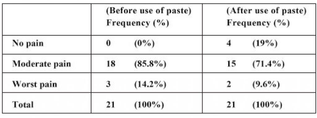
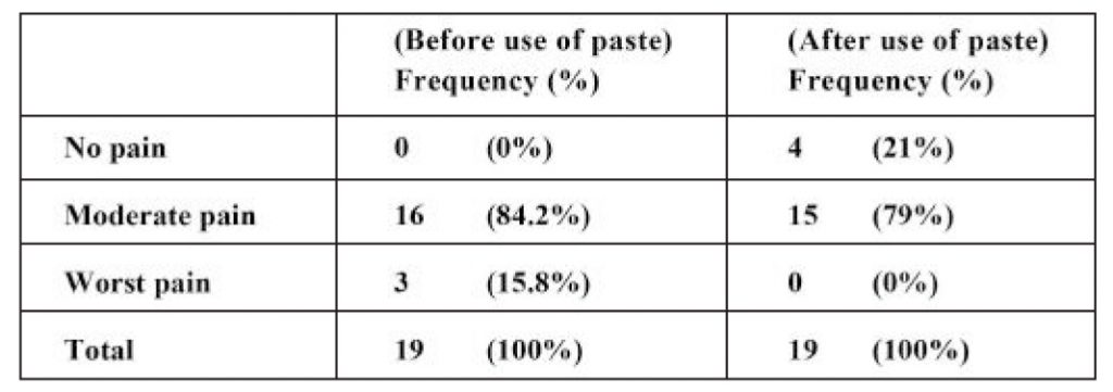
RESULTS: There was greater reduction in moderate pain from 85.8% to 71.4% with strontium chloride and from 84.2% to 79% with potassium nitrate.
CONCLUSION: 10 % w/w strontium chloride is more effective than the 5% w/w potassium nitrate for the management of dentin hypersensitivity.
KEYWORDS: Toothpastes, Sensitivity, Strontium Chloride, Potassium Nitrate
HOW TO CITE: Kiani AZ, Hassan S, Rafiq T. Clinical comparison of 5% potassium nitrate and 10% strontium chloride toothpastes in alleviating dentin hypersensitivity following manual scaling: A pilot study. J Pak Dent Assoc 2021;30(1): 56-60.
DOI: https://doi.org/10.25301/JPDA.301.56
Received: 18 June 2020, Accepted: 14 November 2020

INTRODUCTION

Dentin Hypersensitivity (DH) has been defined as ‘an acute sharp pain of short duration due to exposed dentin, in response to external stimuli that cannot be ascribed to any other form of dental defect and pathology’.1,2 It is a painful response to different irritants such as tooth brushing, sweet and/or sour food etc.3 It is one of the most commonly encountered clinical problems in routine practice. It has also been considered as a chronic pain condition by some authors.4
It is a multi-factorial disease condition caused by enamel loss that could be due to any trauma, gingival recession, erosion and tooth wear.1 The prevalence of DH in the human population is about 10 to 30% , with a female predominance.5 Any surface of tooth can be affected but most common sites are buccal and cervical region of canine and premolars.3 DH can affect any age but usually peaks during the third and fourth decade of life.3
It has been proposed that dentin is composed of dentinal tubules that are covered by mineralized structures on crown by enamel or on the root by the cementum.6 Exposed dentinal tubules due to gingival recession or loss of enamel, play an important role in dentin hypersensitivity.7,8 Many theories have been proposed to explain this phenomenon, including ‘Odontoblast Receptor Theory’, ‘Dentin Innervation Theory’ but the most widely accepted theory is the ‘Hydrodynamic Theory’. According to this theory, when dentinal tubules come into contact with any stimuli, the dentinal fluid present in tubules tends to move towards the pulp. This movement stimulates the nerve endings in the pulp and that leads to discomfort with pain called dentin hypersensitivity.5 Many agents to manage DH are currently being used based on two different mechanisms, either by blockage of nerve activity or tubular occlusion. In tubular occlusion approach, most commonly used agents are stannous, strontium, oxalate salts, arginine, silicas, and bioactive glasses.5
These agents form salt precipitates on the surface of exposed dentin and inside the dentinal tubules. These precipitates effectively reduce or block the fluid movement in the dentinal tubules and exert a desensitization effect.3 The other approach is desensitization of nerve endings, blockage of neural transmission at the pulpal tissues and chemical depolarization of the nerve synapse.4 Most common agent used in this approach is potassium nitrate.4 Ideally, treatment for dentin hypersensitivity should be quick and simple and should be easily available for patients. Thus, the use of specially medicated toothpastes has been adopted as one of the first lines of treatment.7
They are noninvasive and easily available in pharmacies and even in superstores, and according to some studies, they have appreciated cost benefit ratio, in comparison with professional appointments.7 Keeping all of these points in mind, we carried out this pilot study to compare the desensitizing efficacy of two commercially available dentrifices with different active ingredients in a hospital setting in Islamabad, as to our knowledge no such study had been carried out regionally.

METHODOLOGY

This pilot study was based on 40 healthy adults, recruited in the Periodontology department of Islamic International Dental Hospital, Islamabad with the complaint of sensitivity.
The sample size was calculated using the WHO Sample Size Calculator with the aid of the parent articles that were used as a template.9,10 Patients’ sensitivity was confirmed
by using a triple syringe of the dental unit after which manual scaling was done by 3rd year BDS students of Islamic International Dental College. Ethical approval was
given by the Ethical Committee of Riphah International University(Reference Number: IIDC/IRC/2018/04/001). The whole process was explained to the patients and signed consent was obtained before the start of procedure.
Inclusion criteria:
All participants after completing medical history were reviewed by the Principal Investigator before they took part in the study. Participants were selected on the basis of the
following criteria:

  • Both male and female patients
  • Age limit between 18-40 years
  • Subjects with good health (with no known systemic diseases)
  • With Minimum score of 4 on VAS (Visual Analogue Scale)

Exclusion criteria:
Principal Investigator ruled out participants that were:

  • Already using any desensitizing agent
  • Currently going through any dental procedure
  • With any appointment of dental treatment during the next 2 weeks that might affect their hypersensitivity.

Proforma:
The Proforma was based on demographics of participants, checklist for inclusion criteria, procedure done, Visual Analog Scale (VAS)(11) for before and after use with dentifrice, scaling assessment and follow up date. The minimum score on the VAS was “0” and the highest score was set at “10”. VAS was categorized in the following manner.

  • 0 “no pain”
  • 0-6 “moderate pain”
  • 6-10 “worst pain”

Participants were asked to have a look at the VAS and encircle the score given on the scale according to intensity of their sensitivity.
Methodology:
Participants were selected according to our selection criteria. Toothpastes were labeled by the Principal Investigator as “Paste A”, that was Sensodyne Original 50mg with 10% w/w Strontium Chloride, as an active ingredient and, “Paste B” that was Sensodyne Flouride 50mg with main ingredient that was 5% w/w Potassium Nitrate. Operator had no idea about the pastes. Toothpastes were given by lottery method to the participants. Subjects were given written instructions to use the toothpaste twice a day regularly, using a pea sized
amount of toothpaste on medium bristles brush using the Modified Bass brushing technique, which was demonstrated to them on the dental model. All participants were called after two weeks for follow-up. 22 of the patients came back on time for follow-up session, while 18 were contacted via telephone and were sent an image of the VAS and were asked to record their answers. None of the patients were lost to follow-up or dropped out of the study.
Statistical Methodology:
All the data collected after 2 weeks was entered in SPSS Statistics version 17. The mean of the demographics was calculated and tabulated. The MANN WHITNEY test was applied and results were then tabulated and were prepared for the sensitivity before and after the use of the toothpastes. The statistical significance was set at 0.05.

RESULTS

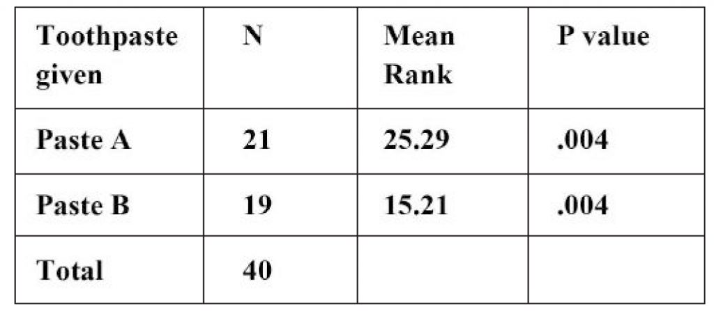
40 Patients were evaluated in our study, 21 subjects were provided with paste A and 19 participants were provided with paste B. Out of 40 subjects, 26 were males (65%) and 14 (35%) were females. Majority of patients were between 20-30 age groups. No side effects were reported in any patients enrolled in this study. (See Table 1)

Table 1: Distribution of pastes among participants

Before commencing the study, 18 patients out of 21 complaint of moderate pain while 3 complaint of worst pain. After use of paste A for the 2 weeks, the 4 patients reported no pain, 15 presented with moderate and 2 with worst pain. (See Table 2)

Table 2: Frequency and percentage of patients before and after use of paste A

At the start of the study, 16 patients out of 19 presented with moderate pain, while 3 presented with worst pain. After use of paste B for the recommended period, 4 patients reported no pain and 15 with moderate pain. No patient reported worst pain. (See Table 3)

Table 3: Frequency and percentage of patients before and after use of paste B

The results obtained were statistically significant as the p value calculated was 0.004. According to the frequencies Paste A performed better than Paste B.

DISCUSSION

Dentin Hypersensitivity (DH), most commonly encountered condition in dental practice, is not only difficult to diagnose but also has an immense effect on the patient’s quality of life.3 In the light of available studies, DH was found to be present in 10% to 30% of the general population.12 A study was conducted which revealed that 42% of DH reported, was in young patients in 18-35 age group.13 Another study showed this peak in third and fourth decade of life with female predominance, affecting mostly premolars and molar teeth.14 In our study mean age group was 20-30 years with generalized sensitivity and with male predominance. Currently a lot of treatment regimens are available with many active ingredients. Most of these agents either work by occlusion of dentinal tubules, for example, Strontium based (chloride, acetate) products or by desensitization of nerve ending, for example, Potassium based (chloride, citrate, nitrate) products.15,16
An in vitro and in vivo study was conducted on rats to check four different toothpastes in occluding exposed dentinal tubules. Potassium nitrate, strontium chloride and potassium
citrate toothpaste containing group were found with less number of open dentinal tubules that was confirmed by the lower permeability of dentin after using desensitizing agents.7
In our study strontium chloride was more effective in occluding dentinal tubules than potassium nitrate Our study compared two commercially available toothpastes containing potassium nitrate and strontium chloride as main active ingredients. Strontium chloride is a protein precipitant and its mechanism of action is the precipitation at the nerve ending and to form a seal to avoid the movement of fluid.3
A study by Minkoff and Axelrod as reported by Porto IC et al stated that regular home use of toothpaste containing 10% strontium chloride proved an efficient means of reducing DH17 which is coherent with our results, showing that 10% strontium chloride is an effective means for management of DH. Kishore et al. evaluated the efficacy of desensitizing
agents and stated that 10% strontium chloride showed a marked reduction in dentinal sensitivity whereas 5% potassium nitrate solution could not.18 Our study shows similar results for 10% strontium chloride and in contrast good results for 5% potassium nitrate as well.
A randomized, double-blind, placebo-controlled study conducted on potassium nitrate, stated that mouthwash containing a mixture of KNO3, NaF, and CPC reduced DH and gingival inflammation.12 Hodosh stated that topical applications of 1-15% potassium nitrate or a paste containing 10% potassium nitrate were effective in minimizing dentin
hypersensitivity.19 In our study, the dentifrice contained 5% potassium nitrate which was also effective to some extent but not so much as 10 % strontium chloride. Increasing the
percentage of potassium nitrate from 5% to 10%, might be helpful for management of DH as suggested in the above study.
Another study conducted on potassium nitrate stated that application of potassium nitrate twice daily for two minutes in the form of dentifrice is better than once daily for two minutes.4 Our study showed same results with application of 5% potassium nitrate twice daily for management of DH.

LIMITATION OF STUDY

This study relied on a proforma only rather than any clinical test to determine dentin hypersensitivity. Our participants were mostly from Islamabad and Rawalpindi; it is not prudent to assume that our findings are applicable to a broader geographic population.

CONCLUSION

Under the limitations of our study, 10% w/w strontium chloride performed adequately in management of dentin hypersensitivity than 5% w/w potassium nitrate. Since this study was conducted in Islamic International Dental Hospital Islamabad and involved a small number of people, a more comprehensive research is required to be done for better results.

SOURCE OF FUNDING

There were no funding sources availed for this study.

CONFLICT OF INTEREST

The authors have stated explicitly that there are no conflicts of interest in connection with this article.

REFERENCES

  1. Alcantara PM, Barroso NFF, Botelho AM, Douglas-de-Oliveira DW, Goncalves PF, Flecha OD. Associated factors to cervical dentin hypersensitivity in adults: a transversal study. BMC Oral Health. 2018;18:155. https://doi.org/10.1186/s12903-018-0616-1
  2. Amit B, Shalu BV. Gingival enlargement induced by anticonvulsants, calcium channel blockers and immunosuppressants: A review. IRJP. 2012;3:116-9.
  3. Majji P, Murthy KR. Clinical efficacy of four interventions in the reduction of dentinal hypersensitivity: A 2-month study. Ind J Dent Res. 2016;27:477-82. https://doi.org/10.4103/0970-9290.195618
  4. James JM, Puranik MP, Sowmya KR. Dentinal Tubule Occluding Effect of Potassium Nitrate in Varied Forms, Frequencies and Duration: An In vitro SEM Analysis. J Clin Diag Res : JCDR. 2017;11:Zc06- zc8.
  5. Ashwini S, Swatika K, Kamala DN. Comparative Evaluation of Desensitizing Efficacy of Dentifrice Containing 5% Fluoro Calcium Phosphosilicate versus 5% Calcium Sodium Phosphosilicate: A Randomized Controlled Clinical Trial. Contemp Clin Dent. 2018;9: 330-36.
  6. Jacobsen PL, Bruce G. Clinical dentin hypersensitivity: understanding the causes and prescribing a treatment. J Contemp Dent Prac. 2001;2:1-12. https://doi.org/10.5005/jcdp-2-1-27
  7. Pinto SC, Silveira CM, Pochapski MT, Pilatt GL, Santos FA. Effect of desensitizing toothpastes on dentin. Braz Oral Res. 2012;26:410-17. https://doi.org/10.1590/S1806-83242012000500006
  8. Lynch MC, Perfekt R, McGuire JA, Milleman J, Gallob J, Amini P, et al. Potassium oxalate mouthrinse reduces dentinal hypersensitivity: A randomized controlled clinical study. J Am Dent Assoc (1939). 2018;149:608-18. https://doi.org/10.1016/j.adaj.2018.02.027
  9. Hall C, Mason S, Cooke J. Exploratory randomised controlled clinical study to evaluate the comparative efficacy of two occluding toothpastes – a 5% calcium sodium phosphosilicate toothpaste and an 8% arginine/calcium carbonate toothpaste – for the longer-term relief of dentine hypersensitivity. J Dent. 2017;60:36-43. https://doi.org/10.1016/j.jdent.2017.02.009
  10. Seong J, Parkinson CP, Davies M, Claydon NCA, West NX. Randomised clinical trial to evaluate changes in dentine tubule occlusion following 4 weeks use of an occluding toothpaste. Clin Oral Investig. 2018;22:225-33. https://doi.org/10.1007/s00784-017-2103-5
  11. Haefeli M, Elfering A. Pain assessment. Europ spine J. 2006;15 Suppl 1:S17-24. https://doi.org/10.1007/s00586-005-1044-x
  12. Hong JY, Lim HC, Herr Y. Effects of a mouthwash containing potassium nitrate, sodium fluoride, and cetylpyridinium chloride on dentin hypersensitivity: A randomized, double-blind, placebo-controlled study. J period Implant Sci. 2016;46:46-56. https://doi.org/10.5051/jpis.2016.46.1.46
  13. West NX, Seong J, Davies M. Management of dentine hypersensitivity: efficacy of professionally and self-administered agents. J Clin Periodontol. 2015;42 Suppl 16:S256-302. https://doi.org/10.1111/jcpe.12336
  14. Rees JS, Addy M. A cross-sectional study of buccal cervical sensitivity in UK general dental practice and a summary review of prevalence studies. Int J Dent Hygiene. 2004;2:64-9. https://doi.org/10.1111/j.1601-5029.2004.00068.x
  15. Karim BF, Gillam DG. The efficacy of strontium and potassium toothpastes in treating dentine hypersensitivity: a systematic review. Int J Dentistry. 2013;2013:573258. https://doi.org/10.1155/2013/573258
  16. Orchardson R, Gillam DG. The efficacy of potassium salts as agents for treating dentin hypersensitivity. J Orofacial Pain. 2000;14:9-19.
  17. Porto IC, Andrade AK, Montes MA. Diagnosis and treatment of dentinal hypersensitivity. J oral science. 2009;51:323-32. https://doi.org/10.2334/josnusd.51.323
  18. Kishore A, Mehrotra KK, Saimbi CS. Effectiveness of desensitizing agents. J Endod. 2002;28:34-5. https://doi.org/10.1097/00004770-200201000-00008
  19. Bartold PM. Dentinal hypersensitivity: a review. Aust Dent J. 2006;51:212-8; 76. https://doi.org/10.1111/j.1834-7819.2006.tb00431.x

1. Registrar, Department of Periodontology, Riphah International University.
2. Assistant Professor, Department of Periodontology, Riphah International University
3. House Officer, Department of Periodontology, Riphah International University.
Corresponding author: “Dr Anam Zahid Kiani” < anamzahid@gmail.com >

Clinical Comparison of 5% Potassium Nitrate and 10% Strontium Chloride Toothpastes in Alleviating Dentin Hypersensitivity Following Manual Scaling: A Pilot Study

Anam Zahid Kiani                       BDS, MSc

Sobia Hassan                               BDS, FCPS

Talha Rafiq                                   BDS

OBJECTIVE: The objective of this study was to compare the efficacy of dentifrices containing potassium nitrate and strontium chloride in reducing dentinal sensitivity pain.
METHODOLOGY: A pilot study was conducted in Islamic International Dental Hospital Islamabad, Pakistan (Duration two weeks). Forty participants were selected, aged between 18-40 years, having dentin hypersensitivity with the minimum score of 4 on Visual Analog Scale. Participants were randomly allocated with dentifrices containing 5% w/w potassium nitrate and 10 % w/w strontium chloride and were asked to use it for 2 weeks and come back for follow up. Statistically significant correlations were found using the Man Whitney test. A pre-designed proforma was used to acquire information about the patient. Sensitivity was determined with the aid of the Visual Analog Scale.
RESULTS: There was greater reduction in moderate pain from 85.8% to 71.4% with strontium chloride and from 84.2% to 79% with potassium nitrate.
CONCLUSION: 10 % w/w strontium chloride is more effective than the 5% w/w potassium nitrate for the management of dentin hypersensitivity.
KEYWORDS: Toothpastes, Sensitivity, Strontium Chloride, Potassium Nitrate
HOW TO CITE: Kiani AZ, Hassan S, Rafiq T. Clinical comparison of 5% potassium nitrate and 10% strontium chloride toothpastes in alleviating dentin hypersensitivity following manual scaling: A pilot study. J Pak Dent Assoc 2021;30(1): 56-60.
DOI: https://doi.org/10.25301/JPDA.301.56
Received: 18 June 2020, Accepted: 14 November 2020
Download PDF

Prevalence of Distal Carious Lesions in Mandibular Second Molars Due to Mesio-Angular Impacted Third Molars

Talha Ashar                                        BDS, FCPS

Asma Shakoor                                   BDS, MSc, MFDSRCS

Sadia Ghazal                                      BSc, M.S

Naghma Parveen                               BDS, MCPS, FCPS

Muhammad Nasir Saleem                BDS, FCPS, MSc

Hina Zafar Raja                                  BDS, FCPS, MSc

OBJECTIVE: This study was conducted to observe the relationship of mesio-angular impacted third molars to the development of distal caries in adjacent second molars.
METHODOLOGY: This cross-sectional study was conducted at Nishtar Institute of Dentistry, Multan. Nine hundred and eighty nine periapical, bitewing and Panoramic radiographs were recorded and examined for distally carious second molars and impacted mandibular third molars. SPSS version 23.0 was used for data entry and analysis.
RESULTS: Almost 40.8% of the target population reported with distal caries due to third molar impactions. A total of 53.30% of these impactions were of the mesio-angular variety. No distal caries was detected in transverse type of impactions.
CONCLUSION: The current study concluded that the prevalence of distal caries in mandibular second molars and the positioning of adjacent impacted mandibular third molars in the dental arch were interlinked. Consequently, extraction of mandibular third molars should be considered to prevent caries and premature loss of second molar teeth.
KEYWORDS: Distal caries, Impacted third molar, Infections, Risk Factor
HOW TO CITE: Ashar T, Shakoor A, Ghazal S, Parveen N, Saleem MN, Raja HZ. Prevalence of distal carious lesions in mandibular second molars due to mesio-angular impacted third molars. J Pak Dent Assoc 2021;30(1):50-55.
DOI: https://doi.org/10.25301/JPDA.301.50
Received: 09 November 2020, Accepted: 17 December 2020

INTRODUCTION

Third molars usually erupt in the oral cavity at the age of 15 to 24 years .Some erupt in the late 30s, while others remain unerupted.1,2 The time of eruption, position in the arch and size of the crown varies among individuals. Third molars are considered as impacted when they fail to erupt into a functional occlusion in the oral cavity at a specified time.3
These impactions are classified according to angulations in the arch, mandibular bone position and crown patterns.3 Pell GJ and Gregory GT devised a comprehensive system to classify impacted wisdom teeth.4Winters categorized the third molars into mesio-angular, disto-angular, horizontal, and vertical impactions – depending on tooth angulation in the oral cavity.5 Previous literature showed a high rate of carious lesions in the second molars due to impaction of third molars.6,7 Majority of the distal caries was linked to mesio-angular tilting of third molar teeth.7,8 Peri-coronal flap makes this area less accessible for routine cleaning and more prone to biofilm and plaque accumulation.9
Gingival recession further accelerates the process by allowing food impaction and exposure of cementoenamel junction. Carious lesions initiate and progress as a consequence.10 Pain is the most significant
indicator in the detection of caries of this variety.11 These lesions often remain unnoticed for long periods of time due to poor visibility. Delay in treatment of distal carries is one of the lead causes of early tooth loss.12
A similar study was carried out in Islamabad, Pakistan with the findings that 42.5% of distal caries in mandibular econd molars developed due to adjacent impacted third molars. A total of 200 patients with impacted third molars were included in the study.13 Another study conducted in Lahore showed the pattern of mandibular third molar impaction and its complications. This study concluded that mesio-angular third molar impaction was most common type of impactions responsible for the occurrence of dental caries in adjacent molars.14 Distal caries in mandibular second molars due to mesially tilted impacted third molars is a tremendous public health concern which needs to be addressed.15 Various studies prove that early extraction of third molars can prevent this; thereby arresting premature tooth loss.16,17 There is a lack of local research on the occurrence of distal caries in mandibular second molar due to third molar impactions. There is a need to observe this occurrence especially within the population visiting dental hospital in Multan. A study was required to know the effects of impaction in geographically diverse area such as Multan. Thus, the rationale of this study was evaluate the relationship of distal caries in mandibular second molars to mesio-angular impacted third molars.
The aims of this study were to;
a) Assess the prevalence of distal caries in mandibular second molars.
b) Assess the relationship between the type of impaction of mandibular third molars and distal caries in adjacent mandibular second molars.

METHODOLOGY

A cross-sectional survey was conducted at Nishtar Institute of Dentistry, Multan for the duration of eight months. An ethical review committee provided the approval for the study (Ref. 9972/NID). Patients were informed of the goals of the study and their consent was taken. All the patients referred for pain in the second molar region were provisionally diagnosed with proximal caries due to third molar impactions.
Concise patient history forms were recorded. After clinical examination, patients aged between 17 to 35 years underwent periapical and bitewing x-rays for assessment and diagnosis of distal caries in mandibular second molars. Panoramic radiographs were used to analyse the angulation of impaction of third molars. A sample of 989 patients – allocated by convenience sampling – was considered for the purpose of this study. A confidence interval of 95% and absolute precision of 5% was established. Taking into account a dropout rate of 10%, the sample was inflated by 10% accordingly. Patients with missing mandibular 2nd molars, systemic diseases and severe periodontitis were excluded from the study.
Mandibular third molars were classified as vertical, mesioangular, distoangular and horizontal according to the Pell GJ and Gregory GT classification system.4 Three clinicians evaluated periapical and bitewing radiographs at different times for detection of distal carries and third molar impactions. An agreement of two or more observers was considered confirmatory for the diagnosis to eliminate any potential bias. It also supported inter examiner reliability.
The study had two variables of interest pertinent to the objectives i.e. “Presence/absence of distal caries” and “Presence/absence of third molar impaction”. Data was analysed using SPSS version 23. Descriptive statistics were applied to assess data in frequencies. Prevalence of distal caries and third molar impactions was displayed in graphs. Mean, standard deviation and percentage values were used to analyse and assess data.

RESULTS

A total of 989 patients exhibiting impacted mandibular third molars were assessed for the purpose of this study. Amongst them, 408 patients (40.8%) were diagnosed with distal caries in mandibular second molars and included in further analysis. Table 1 presents basic data of the study participants. There were more female patients than that of

Table 1: Basic demographics

males. This indicated a higher risk of second mandibular molars distal caries in females. Unilateral impactions were less common than bilateral impactions of mandibular third
molars. Data analysis revealed 66% impactions on both sides of the dental arch with 34% limited to one side only. Mandibular third molars were most commonly impacted in
a mesio-angular dimension. Table no 2 presents patient data segregated according to age groups as follows;

Group 1: 17 to 22 years of age.
Group 2: 23 to 29 years of age.
Group 3: 30 to 35 years of age.
In this study, five different types of mandibular third molar impactions were discussed. Their prevalence and

Table 2: Age Groups and distal caries prevalence

association with distal caries was described in Table no 2. Mesioangular impacted third molars caused the highest incidence of distal caries in second mandibular molars. On the other hand, no caries was observed in transverse impactions.

Figure 1: Gender Distribution

Figure 2: Prevalence of distal caries in different age groups

Figure 3: Distribution of unilateral and bilateral impaction of mandibular third molar

Figure 4: Angulation of impacted 3rd Molar

Figure 5: Prevalence of distal caries

DISCUSSION

An impacted tooth may be defined as a tooth that does not reach the occlusal plane despite two thirds of its root development. Mandibular third molars are the most frequently impacted teeth.18,19 Development of third molar impactions can be attributed to a number of etiological factors.20 These factors include lack of space, retardation of facial growth, late third molar mineralization, distal direction of eruption, early physical maturity, or lack of sufficient eruption force, blockade by physical/mechanical barriers, such as scar tissue, fibromatosis, compact bone, odontogenic cyst, and tumors.20 The prevalence of impacted third molars has been shown to vary in different populations and ethnicities ranging from 9.6% to 68.5%.21,22 These impactions, however, tend to be nearly equally distributed across the two genders in many of the earlier studies with slightly increased frequency in females according to some.22-24 The present study also found similar results with regards to the gender predilection of impacted third molars. There were 54% females and 46 % males with impacted third molars. This can be attributed to the growth pattern differences between the two genders. The growth of mandible in males, continues during the root development of the mandibular third molars giving a chance for these teeth to erupt in the oral cavity. A slightly increased frequency of impacted mandibular third molars in females may be due to an early cessation of jaw growth in this this gender group. This explanation, however, caters to only a single etiological factor of impactions in third molars namely the lack of space.
In the mandible, the impacted third molars tend to occur more frequently bilaterally than unilaterally. This has been shown by some earlier studies23,25,26 and is consistent with our findings of 66% bilateral occurrence versus a 34% unilateral presentation. This can also be explained on the basis of the fact that the local reasons for impacted third molars like lack of space tend to affect both sides in an individual at a certain stage of his/her jaw development. Third molar impactions can be classified in different ways. One of the most commonly employed classification is the Winter classification based on the angle formed by the long axis of the third molar and the second molar.27 This was the classification system used in present study categorizing impactions as mesio-angular, disto-angular, vertical, horizontal and transverse. The most common type of third molar impaction according to our findings was mesio-angular impaction which is in line with most earlier studies from different populations and ethnic backgrounds.23,24 An earlier study conducted in Pakistani population also identified mesio-angular impaction as the most prevalent type of mandibular third molar impaction.28
Impacted third molars have been associated with a variety of different pathological conditions. These conditions include pericoronitis, caries, food lodgment, pocket formation,
periodontal bone loss, root resorption of adjacent teeth, and development of cysts and tumors.28-30 The frequency of dental caries in teeth adjacent to impacted third molars has
been reported to be as low as 1 to 4.7%31,32 to as high as 15-51%18,33-35 in some of the earlier studies. Our study has showed similar trend with 40.8% of distal surface of second
molars affected by dental caries. Very similar results were obtained from an earlier study on Pakistani population exhibiting 42.5% of second molars affected by distal caries in the presence of an impacted third molar.33 Dental caries is a multifactorial disease that can increase in its frequency in individuals with local areas of food impaction. This facilitates the accumulation of dental plaque containing cariogenic bacteria due to the inability of the normal cleaning aids to access these areas.
The highest prevalence of dental caries in our study (56.3%) was associated mesio-angular impaction followed by the horizontal impactions (24.2%). This is in accordance with many other studies displaying similar trend.33,36-38 An earlier study on Pakistani population also exhibited similar trend with mesio-angular and horizontal impactions displaying
41% and 27% distal caries respectively.33 Both mesio angular and horizontal impactions can form plaque accumulating crevices resulting in the distal caries in second molars. A
change in the microbial flora and inflammatory mediators has been proposed by earlier study around impacted third molars strengthening this view point further.37 Cervical caries in surfaces adjacent to the impacted teeth can display carious lesions due to the abnormal contact between the most mesial surface of the impacted third molars and the distal surface of mandibular second molars. The area of contact of impacted third molar with the second molar has also been explored in some earlier studies with contact at the amelocemental junction being the most susceptible.
This is an aspect not covered in our study that can have meaningful clinical implications. Based on the findings of our study, second molar teeth adjacent to mesio angularly and horizontally impacted third molars should be closely either prophylactically removed or followed up closely.

LIMITATIONS

The first limitation of the study was a constrained sample size. This can be overcome by conducting the review on a larger scale with an increased number of patients. However,
this is time consuming and requires a greater number of qualified dentists. The second limitation was the radiographic technique. In the current study, OPG, periapical and bitewing
radiographs were used for assessment and diagnosis. CBCT is a more effective diagnostic tool for presurgical analysis. It eliminates errors of magnification, superimposition and image distortion. Three dimensional imaging allows clear identification of important anatomic landmarks like mandibular canal, mental foramen and maxillary sinus. Additional information such as the thickness of cortical plate, cancellous bone patterns, fenestrations, and roots inclinations can be obtained with the help of CBCT prior to surgical entry.

CONCLUSION

The current study concluded that the prevalence of distal caries in mandibular second molars and the positioning of adjacent impacted mandibular third molars in the dental arch
were interlinked. This relationship provides a favourable environment for the initiation and progression of distal caries in mandibular second molars. Among all types of third molar
impactions, mesio-angular type provides the most favourable space and niche for the colonization of microbes leading to distal caries in the adjacent second molar.

CONFLICT OF INTEREST

The authors declare that there is no conflict of interest. This study was not funded by any organization or institute.

REFERENCES

  1. Ventä I, Schou S. Accuracy of the Third Molar Eruption Predictor in predicting eruption. Oral Surg Oral Med Oral Pathol Oral Radiol Endod. 2001; 91:638-42. https://doi.org/10.1067/moe.2001.113350
  2. Kruger E, Murray W, Thomson, MA, Konthasinghe P. Third molar outcomes from age 18 to 26: Findings from a population-based New Zealand longitudinal study. Oral Surg Oral Med Oral Pathol Oral Radiol Endod. 2001; 92:150-55. https://doi.org/10.1067/moe.2001.115461
  3. Breik O, Grubor D. The incidence of mandibular third molar impaction in different skeletal face types. Aust Dent J. 2008; 53:320-24. https://doi.org/10.1111/j.1834-7819.2008.00073.x
  4. Pell GJ, Gregory GT. Impacted mandibular third molars: Classification and modified technique for removal. The Dental Digest. 1933; 39:330-38.
  5. Obimakinde, OS. Impacted mandibular third molar surgery; an overview. A publication by the dentiscope editorial Board .2009; 16:22-24.
  6. Faculty of Dental Surgery of the Royal College of Surgeons of England FDS response to NICE review 2014 of guidance on the extraction of wisdom teeth. 2015. Online information available at https://www.rcseng.ac.uk/fds/policy/documents/nice-reviewof guidanceon-wisdom-teeth-extraction-fds-response-with-appendices/view (accessed March 2015).
  7. McArdle LW, Renton TF. Distal cervical caries in the mandibular second molar: an indication for the prophylactic removal of the third molar? Brit J Oral Maxillofac Surg 2006;44:42-5. https://doi.org/10.1016/j.bjoms.2005.07.025
  8. Toedtling V, Yates JM. Revolution vs status quo? Non-intervention strategy of asymptomatic third molars cause harm. Brit Dent J. 2015; 219: 11-12. https://doi.org/10.1038/sj.bdj.2015.525
  9. Chu FC, Li TK, Lui VK, Newsome PR, Chow RL, Cheung LK. Prevalence of impacted teeth and associated pathologies- A radiographic study of the Hong Kong Chinese population. Hong Kong Med J. 2003; 9:158-63.
  10. BDA evidence summary. Second molar distal caries; 2013 (cited June 2013). Available from http://www.bda.org/dentists
  11. Mansoor J, Jowett A, Coulthard P. ‘NICE or not so NICE?’ Brit Dent J. 2013; 215: 209-212.https://doi.org/10.1038/sj.bdj.2013.832
  12. Krishnan B, Mohammad Hossni El Sheikh, Rafa El-Gehani, Orafi H. Indications for removal of impacted mandibular third molars: A single institutional experience in Libya. J Maxillofac Oral Surg. 2009; 8:246-48. https://doi.org/10.1007/s12663-009-0060-5
  13. Sheikh AZ,Riaz M,Shafiq S. Incidence of distal caries in mandibular second molars due to impacted third molars – A clinical & radiographic study. Pak Oral Dent J.2012;32:364-70.
  14. Rauf S, Ali W, Chaudhry R, Kazmi SS, Imtiaz M. Pattern of mandibular third molar impaction: a radiographic study. Pak Oral Dent J 2019; 39:238-42.
  15. American Association of Oral and Maxillofacial Surgeons (AAOMS). Age-Related Third Molar Study. J Oral Maxillofac Surg 2005; 63: 1106-114. https://doi.org/10.1016/j.joms.2005.04.022
  16. American Association of Oral and Maxillofacial Surgeons (AAOMS) Conventional Wisdom about Wisdom Teeth Confirmed: Evidence shows keeping wisdom teeth may be more harmful than previously thought. 2010. Online information available at http://www.aaoms.org/ (accessed May 2015).
  17.  Chu FC, Li TK, Lui VK, Newsome PR, Chow RL, Cheung LK.Prevalence of impacted teeth and associated pathologies- A radiographi study of the Hong Kong Chinese population. Hong Kong Med J. 2003; 9:158-63.
  18. van der Linden W, Cleaton-Jones P, Lownie M. Diseases and lesions associated with third molars: Review of 1001 cases. Surg, Oral Med, Oral Pathol, Oral Radiol, Endodontol. 1995;79: 142-45. https://doi.org/10.1016/S1079-2104(05)80270-7
  19. Bishara SE, Andreasen G. Third molars: a review. American J Orthod. 1983;83:131-37. https://doi.org/10.1016/S0002-9416(83)90298-1
  20. Haidar Z, Shalhoub SY. The incidence of impacted wisdom teeth in a Saudi community. Int J Oral maxillofac Surg. 1986;15:569-71. https://doi.org/10.1016/S0300-9785(86)80060-6
  21. Hatem M, Bugaighis I, Taher EM. Pattern of third molar impaction in Libyan population: A retrospective radiographic study. The Saudi J Dent Res. 2016;7:7-12. https://doi.org/10.1016/j.sjdr.2015.04.005
  22. Quek S, Tay C, Tay K, Toh S, Lim K. Pattern of third molar impaction in a Singapore Chinese population: a retrospective radiographic survey. Int J Oral and Maxillofac Surg. 2003;32:548-52. https://doi.org/10.1016/S0901-5027(03)90413-9
  23. Rezaei F, Imani MM, Khavid A, Nabavi A. Patterns of Mandibular Third Molar Impaction in an Iranian Subpopulation. Pesquisa Brasileira em Odontopediatria e Clínica Integrada. 2020;20:5411. https://doi.org/10.1590/pboci.2020.099
  24. Eshghpour M, Nezadi A, Moradi A, Shamsabadi RM, Rezaei NM, Nejat A. Pattern of mandibular third molar impaction: A cross-sectional study in northeast of Iran. Niger J Clin Pract. 2014;17:673-7. https://doi.org/10.4103/1119-3077.144376
  25. Guthua S, Mwaniki D. A retrospective study of characteristics of impacted mandibular wisdom teeth in 110 patients treated in Nairobi, Kenya. African dental journal: official publication of the Federation of African Dental Associations J dent Africa. 1992;6:30-3.
  26. Ramamurthy A, Pradha J, Jeeva S, Jeddy N, Sunitha J, Kumar S. Prevalence of mandibular third molar impaction and agenesis: a radiographic south Indian study. J Ind Acad Med Radiol. 2012;24:7. https://doi.org/10.5005/jp-journals-10011-1289
  27. Winter GB. Impacted mandibular third molar. St Louis: American Medical Book. 1926;41.
  28. Ishfaq M, Wahid A, Rahim AU, Munim A. Patterns and presentations of impacted mandibular third molars subjected to removal at Khyber college of dentistry Peshawar, Pak. Oral Dent J. 2006;26:221-26.
  29. Al-Anqudi SM, Al-Sudairy S, Al-Hosni A, Al-Maniri A. Prevalence and Pattern of Third Molar Impaction: A retrospective study of radiographs in Oman. Sultan Qaboos University Medi J. 2014;14:e388.
  30. Stanley H, Alattar M, Collett W, Stringfellow Jr H, Spiegel E. Pathological sequelae of “neglected” impacted third molars. J Oral Pathol Med. 1988;17:113-17.
    https://doi.org/10.1111/j.1600-0714.1988.tb01896.x
  31.  Daley TD. Third molar prophylactic extraction: a review and analysis of the literature. Gen Dent. 1996;44:310.
  32. Song F, O Meara S, Wilson P, Golder S, Kleijnen J. The effectiveness and cost-effectiveness of prophylactic removal of wisdom teeth: NHS Centre for Reviews and Dissemination; 2000. https://doi.org/10.3310/hta4150
  33. Sheikh MA, Riaz M, Shafiq S. Incidence of distal caries in mandibular second molars due to impacted third molars-A clinical and radiographic study. Pak Oral Dent J. 2012;32t.
  34. Oderinu OH, Adeyemo WL, Adeyemi MO, Nwathor O, Adeyemi MF. Distal cervical caries in second molars associated with impacted mandibular third molars: a case-control study. Elsevier; 2012. https://doi.org/10.1016/j.oooo.2012.03.039
  35. Rahman NA, Daud MKM, Yaacob MF, Yusoff A. Mandibular Third Molar Impaction and Dental Caries among Patients Attending Hospital Universiti Sains Mslaysia (HUSM). Int Medi J. 2009;16:53-6.
  36. Knutsson K, Brehmer B, Lysell L, Rohlin M. Pathoses associated with mandibular third molars subjected to removal. Oral Surg, Oral Med, Oral Pathol, Oral Radiol, Endodontol. 1996;82:10-7. https://doi.org/10.1016/S1079-2104(96)80371-4
  37. Blondeau F, Daniel NG. Extraction of impacted mandibular third molars: postoperative complications and their risk factors. J Canadian Dent Assoc. 2007;73:325-325e
  38. Sursala M, Blaeser B, Magalnick D. Third molar surgery and associated complication. Oral Maxillofacial Surg Clin North Am. 2003;15:177-86. https://doi.org/10.1016/S1042-3699(02)00102-4

  1. Assistant Professor, Department of Operative Dentistry, Nishtar Institute of Dentistry, Multan, Pakistan.
  2. Associate Professor, Department of Community & Preventive Dentistry, Institute of Dentistry, CMH Lahore Medical College
  3. Dental Technologist, Tehsil Headquarter Hospital Mian Channo.
  4. Associate Professor, Department of Operative Dentistry, Nishtar Institute of Dentistry, Multan, Pakistan.
  5. Professor, Department of Operative Dentistry, Institute of Dentistry, CMH Lahore Medical College, Lahore, Pakistan.
  6. Professor, Department of Prosthodontics, Institute of Dentistry, CMH Lahore Medical College, Lahore, Pakistan.
    Corresponding author: “Dr. Asma Shakoor” < asmashakoor@hotmail.com >